Circulation
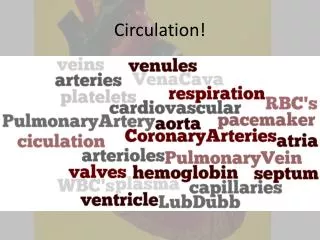
Circulation. What is Circulation?. Tranpsort Exchange must take place at cellular level All cells must have access to an aqueous environment Unicellular vs Multicellular Circulation connects aqueous environment of cell to exchange organs Gases, absorb nutrients, & dispose of wastes.

Circulation
E N D
Presentation Transcript
What is Circulation? • Tranpsort • Exchange must take place at cellular level • All cells must have access to an aqueous environment • Unicellular vs Multicellular • Circulation connects aqueous environment of cell to exchange organs • Gases, absorb nutrients, & dispose of wastes
Simple Circulation • GVC – no true circulation system • Cnidarians - Digest & distributeInner & outer layers bathed in fluids
Advanced Circulatory Systems • 3 basic components of Circulatory System • Circulatory Fluid • Set of Tubes • Muscular Pump • Pump forces fluid through tubes • Muscular heart pumps specialized fluid (such as blood) through tubular vessels • Blood or blood alternative • Carries O2 and nutrients to body tissues • Carries away CO2 and wastes
Open Circulatory System • Arthropods & Molluscs • No distinction between blood & interstitial fluid • HEMOLYMPH • Interconnected system of sinuses & spaces around organs for exchange • Heart is dorsal, elongated • Less energy to build & maintain; but Less effective, slowest M.R.
Closed Circulatory System • Earthworms, Cephalopods, & Vertebrates • Blood confined to vessels & separate from Interstitial Fluid • 1 or more hearts pump into large vessels that branch into smaller ones through organs • Exchange occurs between IF & Blood • High energy cost; more effective & supports higher metabolism
Focus on Vertebrate Circulation • Evolved from a heart w/ 2 chambers, 1 circuit to a double heart w/ 2 circuits • pulmonary and systemic • Vertebrate heart 1 or 2 atria & 1 or 2 ventricles • Vessels • Arteries – carry away; smallest unit - Arteriole • Capillaries – porous exchange points; arranged in beds • Veins – return to heart; smallest unit - Venule • Higher Metabolic Rate = More Complex Cardiovascular rate
Mammals and Birds • Complete division of R& L sides • Left receives & pumps only O2 rich • Right receives & pumps O2 poor • Supports endothermy • 10x as much energy & food as ectothermy
Mammalian Heart • A four-chambered pump • Two atria at top • Two ventricles at bottom • Atrioventricular (AV) valves • Semilunar (SL) valves • Blood is pumped into two separate circuits • Pulmonary circuit (right heart) • Systemic circuit (left heart
Mammalian Heart - Valves • Connective tissue that prevents backflow & keep blood moving 1-way • Control blood flow leaving heart; 1 way doors • Forced open by pressure generated by ventricle • Close when chamber is relaxed
Cardiac Cycle • Systole–diastole sequence is the cardiac cycle • Systolic pressure • Contraction of ventricles pushes blood into arteries at peak pressure • Diastolic pressure • Between contractions, blood pressure in arteries falls to a minimum pressure • Neurogenic hearts (in some crustaceans) • Beat under control of nervous system • Myogenic hearts (all other animals) • Contractions initiated within the heart
Cardiac Cycle • Contraction of atria is initiated by signals from the sinoatrial (SA) node (pacemaker cells) • Contraction of ventricles follows, through excitation of atrioventricular (AV) node • Via Purkinje fibers • Electrocardiogram (ECG/EKG)
Blood Vessels • Arteries carry blood away from the heart • Arterioles (small branches of arteries) deliver blood to capillaries • Capillaries exchange material with interstitial fluid • Venules collect blood from capillaries • Veins return blood to the heart
Blood Vessels • Blood moves fastest Aorta & slowest in Capillaries • Blood Pressure – force of pressure against walls of blood vessels • ~120 mm Hg • Emptying of arteries between heartbeats is the diastolic pressure • ~80 mm Hg
Arteries • Artery walls • Inner endothelial layer • Middle layer of smooth muscle • Outer layer of elastic fibers • Arterioles (smallest arteries) constrict and dilate • Regulate flow and pressure of blood into capillaries
Capillaries • Capillary walls • Single layer of endothelial cells • Variation in contraction of smooth muscles of arterioles & pre-capillary sphincters controls blood flow through capillaries • Capillaries of brain, heart, kidneys, liver always filled to capacity; other sites vary over time • After eating blood to Sm. Intestine • Exercise blood directed to muscles & skin • Rings of smooth muscle – pre-capillary sphincters • Contraction in arteriole constricts vessel reducing blood flow • Controls blood flow between arteries & veins
Exchange Across Capillary Walls • Rate of blood flow is slower to maximizes exchange time • Blood traveling through capillaries delivers nutrients, oxygen, and removes wastes from cells • Two major mechanisms drive exchange of substances • Diffusion along concentration gradients • Bulk flow • At Arteriole end Blood pressure is greater than Osmotic Pressure • Pushes Capillary fluid out to cells – oxygen & nutrients • At Venous end Osmotic Pressure is greater than Blood Pressure • Fluid returns back into capillaries from cells – carbon dioxide & wastes
Veins • Veins have thinner walls than arteries • Allows vessels to expand and contract • Veins act as blood reservoirs as well as conduits • Pressure from movements of skeletal muscles and respiration help return blood to heart • One-way valves prevent blood from flowing backward
The Lymphatic System • Extensive network of vessels • Collects excess interstitial fluid (becomes lymph) • Returns it to the venous blood • Lymph nodes, spleen, thymus, tonsils • Remove viruses, bacteria, damaged cells, and cellular debris from lymph and bloodstream • Defend the body against infection and cancer • Returned to blood via lymphatic system • Fluid enters through lymph capillaries intertwined w/ blood capillaries • Drains back into blood @ Vena Cava • Lymph vessels resemble veins & use valves & muscle contraction to move
Maintaining Blood Flow and Pressure • Regulated by controlling • Cardiac output • Degree of constriction of blood vessels (arterioles) • Total blood volume • Autonomic nervous system and endocrine system interact to coordinate these mechanisms
Regulation of Cardiac Output • Baroreceptors detect blood pressure changes and send signals to the medulla • Brain stem sends signals (autonomic nervous system) that alter rate and force of heartbeat • Hormones secreted by several glands regulate both cardiac output and arteriole diameter • Epinephrine & norepinephrine
Local Controls Regulate Arteriole Diameter • Low O2 and high CO2 concentrations in tissues dilate arteriole walls • Increasing arteriole diameter and blood flow • High O2 and low CO2 concentrations have opposite effects • NO released by arterial endothelial cells increase arteriole diameter and blood flow
Mammlian Blood • Fluid connective tissue • Blood cells (erythrocytes, leukocytes, platelets) • Suspended in a fluid matrix (plasma) • Contains water, ions, dissolved gases (O2 and CO2), glucose, amino acids, lipids, vitamins, hormones, and plasma proteins • Plasma proteins • Albumins (transport, osmotic balance, pH) • Globulins (transport, immunoglobins) • Fibrinogen (blood clotting)
Blood & its Parts • RBC’s – erythrocytes • Contain hemoglobin (transports O2 from lungs to body) • 5-6 million per microliter; 25 trillion per 5 L of blood • No nuclei so more room for hemoglobin • WBC’s – Leukocytes • Defend body against infecting pathogens • monocytes, neutrophils, basophils, eosinophils, & lymphocytes • Patrol Interstitial Fluid & Lymph • Platelets • Functional cell fragments that trigger clotting • Platelets – fragments of cells; no nuclei • Originate as pinched off cytoplasmic fragments of larger cells in bone marrow • Function in blood clotting; self-sealing material that plugs leaks • Inactive fibrinogen fibrin
Blood Stem Cells & Cellular Elements • RBCs last 3-4 months • Destroyed in liver & spleen • All blood cells develop from common source w/in bone marrow • Pluripotent stem cells • any type of BLOOD cell • EPO – naturally stimulates production of RBCs • Produced by kidney
Cardiovascular Disease • Cholesterol • LDL – deposits plaque in arteries • HDL – reduces deposition • Exercise HDL’s • Atherosclerosis • Thickening of arteries • Interferes w/ circulation • Hypertension • BP because of narrowing of vessels by atherosclerosis or reduced elasticity • Threat of heart attack or stroke increases with these diseases • Heart Attack – death of cardiac muscle • Stroke – death of nervous tissue in brain • Thrombus – blood clots or clogs in arteries
Gas Exchange • Process by which animals exchange O2 and CO2 with the environment • Two primary operating features of gas exchange • The respiratory medium, either air or water • The respiratory surface, a wetted epithelium over which gas exchange takes place
Respiratory Surface – Tracheal Systems • Some invertebrates, skin is the respiratory surface • Others & all vertebrates, gills or lungs are the primary respiratory surfaces • Simple diffusion of molecules drives exchange of gases across the respiratory surface • Area of respiratory surface determines total quantity of gases exchanged by diffusion • Concentration gradients of O2 and CO2 across respiratory surfaces are kept at optimal levels by ventilation and perfusion (capillaries)
Water Breathers • Respiratory surfaces are wetted by direct exposure to the environment • Require significant energy to keep respiratory surfaces ventilated • High density & viscosity of water • Relatively low O2 content compared with air • Sponges, Cnidarians, Flatworms • Use outer cells to form respiratory surface
Gills (Water Breathers) • Evaginations of the body surface • Water moves over the gills • By the beating of cilia • Is pumped over gills by contractions of body muscles • In sharks, bony fishes, and some crabs • Water moves in a one-way direction over the gills • Maximizes exchange of gases over the respiratory surface
Air Breathers • Air is high in O2 content • Allows air-breathers to maintain higher metabolic levels than water breathers • Air has lower density and viscosity than water • Allows air breathers to ventilate respiratory surfaces with relatively little energy
Insects: Tracheal System • Insects breathe by a tracheal system • Air-conducting tubes (trachea) lead from the body surface (through spiracles) and branch to all body cells • Gas exchange takes place in fluid-filled tips at ends of branches
Respiratory Surfaces - Lungs • Invaginations of the body surface • Allow air to become saturated with water before it reaches the respiratory surface • Reduce water loss by evaporation • Amphibians – small lungs; also use skin • Reptiles, birds, mammals – larger lungs • Size & complexity correlate w/ Metabolic Rate
Lung Ventilation • Positive pressure breathing • Air is forced into lungs by muscle contractions • Negative pressure breathing • Muscle contractions expand lungs, lowering air pressure inside • Allows air to be pulled into the lungs
Mammalian Respiratory System • Air enters the respiratory system through the nose and mouth and passes through the pharynx, larynx, and trachea • Trachea divides into two bronchi leading to lungs • Within lungs, bronchi branch into bronchioles, leading into alveoli surrounded by networks of blood capillaries
Mammalian Respiratory System • Negative pressure mechanism • Air is exhaled passively • Relaxation of diaphragm and external intercostal muscles between ribs • Elastic recoil of lungs (pleural membranes) • Deep and rapid breathing • Forceful expulsion of air driven by contraction of internal intercostalmuscles • Atmospheric pressure = 760 mm Hg • Oxygen in atmosphere is 21% = .21 x 760 = 160 mm Hg • Gas always diffuses from higher partial pressure to lower partial pressure • Lungs must be lower so oxygen moves in
Ventilating Lungs • Tidal volume • Amount of air moved in and out of lungs during an inhalation and exhalation • Vital capacity • Total volume of air a person can inhale and exhale by breathing as deeply as possible • Residual volume • Air remaining in the lungs after as much air as possible is exhaled
Breathing Control • Control mechanisms • Local chemical controls • Regulation centers in the brain stem • Control functions • Match rate of air and blood flow in lungs • Link rate and depth of breathing to body’s requirements for O2 uptake and CO2 release • Sensory receptors in medulla detect changes in levels of O2 and CO2 in blood and body fluids • Carotid bodies • Aortic bodies
Monitoring the blood • CO2 concentration – Carbonic acid – pH sensor in arteries • pH, CO2 so breath deeper breaths • O2 levels measured directly • O2, increase breathing rate • Hyperventilating • Rapid, deep breathes caused by release of too much CO2 • Breathing control center stops signaling ribs & diaphragm • Restored when CO2 or O2 • CO2 during exercise Breathing Rate to removal of CO2
Respiratory Pigments • Oxygen has low solubility in blood; so carried by pigments • Hemocyanin – arthropods & molluscs – uses copper as transfer element • Bluish blood • Hemoglobin – vertebrates & some inverts – uses iron is transfer element • Binding of Oxygen to hemoglobin – Bright Red • W/out Oxygen hemoglobin – dark-purplish Red