Anatomy of the Eyeball and Visual Structures
Comprehensive overview of the anatomy and structures of the eyeball, covering layers, muscles, nerves, and functions. Includes detailed descriptions and functions of each component.

Anatomy of the Eyeball and Visual Structures
E N D
Presentation Transcript
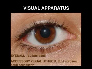
VISUAL APPARATUS EYEBALL - bulbus oculi ACCESSORY VISUAL STRUCTURES - organa oculi accessoria
Eyeball(AP diameter 24 - 26 mm) MRL Layers: 1. tunica fibrosa 2. t. vasculosa 3. t. interna polus ant. fovea centralis linea visus (visual axis) polus post. seen object axis bulbi ext. n. opticus MRM
■fibrous skeleton of the eyeball ■attachment of muscles 1. tunica fibrosa Sclera - post. 5/6 ■ limbus sclerae ■ sulcus sclerae ■ sinus ven. sclerae ■l. cribrosa sclerae Cornea - ant. 1/6 ■limbus corneae ■facies ant. et post.
2. tunica vasculosa Choroid (2/3) supplies the retina pigmented – light-absorbing vascular Corpus ciliare attachment for the lens controls thickness of the lens secretion of aqueous humor Iris contractile diaphragm with a central aperture – pupil ■facies ant. et post. ■ sphincter, dilator Processus ciliares
M. ciliarisAccommodation of the lens increased convexity of the lens – refraction suitable for near vision Innervation: E-W ncl. CN III
M. sphincter pupillaeMiosis M. dilatator pupillaeMydriasis E-W ncl. CN III Symp.ncl.
3. tunica interna ■ pars optica (rods, cones) Retina ora serrata ■ pars caeca: p. iridica p. ciliaris
Layers of the retina N. opticus str. ganglionare n. optici str. ganglionare retinae str. neuroepitheliale str. pigmentosum
Photoreceptors: • Rods • 120 million • primary source of visual information at night • used in peripheral vision • Cones • 7 million • detection of color • central fovea – responsible for central vision
Ophthalmoscopy Fundus - OCULA DEXTRA (OD) Discus (papilla) n. optici Macula lutea + fovea centralis- the area of most acute vision
A. et v. centralis retinae 1a,b a. et v. temporales 2a,b a. et v. nasales 1b 2b 1a 2a
Chambers - camera bulbi ant. et post. Aqueous humor - humor aquosus Sinus venosus sclerae (canal of Schlemm) A P Fibrae zonulares
Lens facies ant. facies post. Optical power + 10 - 17 dioptries axis 4 mm polus ant. polus post. ■ capsula lentis ■cortex lentis ■nucleus lentis aequator
Cataracts loss of transparency cloudiness ageing process
Vitreous chamber: vitreous humor, vitreous body jelly-like substance with watery fluid holds the retina in place supports the lens
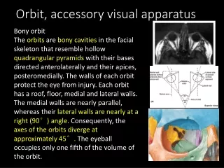
Extraocular muscles Non-striated ■ m. orbitalis (symp.) Palsy =Enophtalmus ■mm. tarsales (symp.) =Ptosis Striated ■m. rectus bulbi sup., inf., med., lat. ■m. obliguus sup., inf. ■m. levator palp. sup.
Anulus tendineus communis
m. rectus sup. (n. III.) m. obliguus sup. (n. IV.) L M m. rectus med. (n. III.) m. rectus lat. (n. VI.) m. obliguus inf. (n. III.) m. rectus inf. (n. III.)
Eyelids - palpebrae Conjunctiva ■palpebra sup. et inf. ■commisurae palpebr. ■rima palpebrarum ■angulus oculi med. et lat. ■facies ant. et post.
1 2 3 3 2 1 Structure of the eyelid 1septum orbitale 2tarsus sup. et inf. 3 lig. palp. med. et lat.
■ skin ■m. orbicularis oculi ■septum orbitale + tarsi ■conjunctiva gll. tarsales f. ant. f. post. cilia gll. ciliares gll. sebaceae limbus palpebr. ant. limbus palpebr. post. Rivus lacrimalis
Chalazion (tarsal chalazion) Hordeolum (sty)
Tunica conjunctiva ■tun. conj. palpebrarum ■tun. conj. bulbi
Apparatus lacrimalis Glandula lacrimalis > ductuli excretorii >lacus lacrimalis ■papillae lacrimales ■puncta lacrimalia ■canaliculi lacrimales ■saccus lacrimalis ■ductus nasolacrimalis
1 2 3 3 2 1 Pars orbitalis Pars palpebralis
Angulus medialis: ■ lacus lacrimalis■caruncula lacrimalis ■plica semilun. conjunctivae
Blood supply A. opthalmica V ophtalmica sup. V. ophtalmica inf. aa. ciliares ant. aa. conjunctiv. v. vorticosa aa. episclerales aa. ciliares post. breves et longae a. centralis retinae
Illustrations and photographs were copied from: Atlas der Anatomie des Menschen/Sobotta. Putz,R., und Pabst,R. 20. Auflage. München: Urban & Schwarzenberg, 1993 Netter: Interactive Atlas of Human Anatomy. Windows Version 2.0