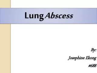
Lung Abscess
Lung Abscess. By: Josephine Ekong #688. CONTENTS. Definition. Classification. Microbiology. Pathophysiology. Clinical manifestation. Diagnosis. Differential Diagnosis. Treatment. DEFINITION.

Lung Abscess
E N D
Presentation Transcript
LungAbscess By: Josephine Ekong #688
CONTENTS Definition Classification Microbiology Pathophysiology Clinical manifestation Diagnosis Differential Diagnosis Treatment
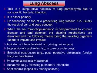
DEFINITION Lung abscess is defined as necrosis of the pulmonary tissue and formation of cavities containing necrotic debris or fluid caused by microbial infection. Localized collection of pus within the lung parenchyma. • Microbial infection causes suppurative necrosis of the lung parenchyma producing a cavity. Often communicate with large airways resulting in cough, expectoration of purulent sputum and air-fluid level on imaging studies .
Lung abscess is caused by: • Bronchial obstruction eg. Cancer • Aspiration of oropharyngeal contents (especially in patients predisposed to loss of consciousness eg. Alcoholics, epileptics ) • More common in pre-abx era with progression bacterial pneumonia +/- empyema
CLASSIFICATION Classification is based on: Duration of symptoms prior to diagnosis; We have the • Acute < 1month • Chronic > 1 month
Primary lung abscess: • used when abscess develops in individuals prone to aspiration or in general good health • 80% of lung abscess is primary (50% of these associated with putrid sputum) • Secondary lung abscess: • obstructive airway neoplasm as a complication of intra-thoracic surgery or systemic condition/treatment that compromises host immune defense mechanisms We also have: Primary lung abscess Secondary lung abscess
MICROBIOLOGY INFECTIOUS ORGANISMS • Bacteria (most common) • Usually: mouth floraanaerobes, most frequently isolated anaerobes: • Peptostreptococcus, • Fusobacteriumnucleatum, • Prevotellamelaningogenica • Bacteroides • Less common: • Staphylococcus aureus, Streptococcus pyogenes, • Pseudomonas aeruginosa, Klebsiellapneumoniae, • Streptococcus pneumoniae • gram-negative bacilli, such as E. coli , Haemophilusinfluenzae type B • Legionella, Nocardiaasteroides
Mycobacteria • M. tuberculosis, M. avium complex, • M. kansasii, other mycobacteria • Fungi • Aspergillus spp., Histoplasmacapsulatum, • Pneumocystiscarinii, Coccidioidesimmitis, • Blastocystishominis ,cryptococcus • Parasites • Entamoebahistolytica, • Paragonimuswestermani, • Strongyloidesstercoralis (post-obstructive)
Noninfectious Causes • Neoplasms Primary lung cancer, metastatic carcinoma, lymphoma • Pulmonary infarction Due to bland embolus (may be secondarily infected in <5%) • Septic embolism Tricuspid endocarditis due to S. aureus and others (typically with positive blood cultures), jugular venous septic phlebitis due to Fusobacteriumnecrophorum (Lemierre syndrome)
Noninfectious Causes • Vasculitis: Wegener's granulomatosis, rheumatoid lung nodule • Airway disease: Bullae, blebs, or cystic bronchiectasis (usually thin-walled) • Developmental: Pulmonary sequestration • Others include: Sarcoidosis, transdiaphragmatic bowel herniation giving appearance of cavity with air-fluid level
PATHOPHYSIOLOGY • Most occur as a complication of aspiration pneumonia and are polymicrobial due to anaerobic bacteria that are normal oral flora • Initial aspiration lung insult may be due to direct chemical injury from aspirated stomach acid or to areas of obstruction due to aspirated particulate matter (i.e. food) • Secondary bacterial infection then may occur.
If the bacterial inoculum is large/virulent or lung defense mechanisms compromised, infection can occur without prior insult to the lung . • Studies of patients with known time of aspiration suggest that tissue necrosis with lung abscess formation takes at least 1 week and up to 2 weeks to develop.
Aspirated bacteria are carried by gravity to dependent portions of the lung. • Due to bronchus/carina anatomy, occur most frequently in posterior segment of RUL then posterior segment of LUL and then the superior segments of RUL/LLL • 1/3 develop empyema due to direct extension . • Amebic lung abscess typically occurs in RLL due to direct extension of liver abscess through the diaphragm
Airway Obstruction • Neoplasm: 50% of lung abscesses in patients >50 are associated with lung carcinoma (post-obstruction vs. necrotic tumor) • Foreign bodies, extrinsic compression (enlarged lymph node • Patients with cell-mediated immunity (AIDS, transplant): OI pathogens such as mycobacteria, Nocardia, Aspergillus, Rhodococccus • Neutropenic patients: • P. aeruginosa, S. aureus, fungi (Aspergillus, Zygomycetes)
Other processes: • bronchiectasis, • secondary infection from bland pulmonary infarction/PE, • septic emboli from TV endocarditis • (involve multiple, noncontiguous areas of the lung), • suppurative phlebitis • Lemierre syndrome: septic phlebitis of the neck (Fusobacterium) with embolic infection in the lung may complicate oropharyngeal infection (peritonsillar abscess)
Histology of lung abscessshows dense inflammatory reaction (low power)
Histology of lung abscess shows dense inflammatory reaction (high power)
diagnosis • *Clinical features Radiological diagnosis Procedures LABORATORY STUDIES
Clinical features Symptoms • The onset may be abrupt or gradual. • Symptoms include fever, sweating, cough and chest pain simulating pneumonia. • The cough is often non productive at first or may produce mucoid or mucopurulentexpectorate from bronchial inflammation close to the abscess area and sometimes there is blood streaking.
There is an expectoration of foul-smelling brown or gray sputum (in anaerobic organisms) or green or yellow sputum • If this happens suddenly in large quantities, this denotes a rupture of the abscess cavity into the bronchus and blood streaking is common. • Pleuritic chest pain, especially with coughing is common because the abscess is near to the pleura. • Weight loss, anaemia, and clubbing or pulmonary osteoarthropathy appear when the abscess becomes chronic (8-12 weeks after onset).
Signs • May be minimal. Consolidation due to pneumonia surrounding the abscess is the most frequent finding. • Inspiratoryrales and pleural rub may be heard. • Rarely the amphoric or cavernous breath sounds are heard • Rupture into the pleural space produces signs of effusion or hydro pneumothorax. • Digital clubbing may develop rapidly.
Amebic lung abscess Patients who develop an amebic lung abscess often have symptoms associated with a liver abscess. These may include right upper quadrant pain and fever. • After perforation of the liver abscess into the lung, the patient may develop a cough and expectorate a chocolate or anchovy paste–like sputum that has no odor.
B) Radiological diagnosis • Lung abscesses as a result of aspiration most frequently • occur in the posterior segments of the upper lobes or the • superior segments of the lower lobes. • A dense shadow is the initial finding before rupture. • Later this opacity develops a cavity with fluid level.may have smooth and regular walls. • Chest films may also reveal associated primary lesions, • e.g., a bronchogenic carcinoma. • malignant abcessshows eccentericcavitation with thick rough irregular walls. • Low-attenuation central region and rim enhancement on • CT scan
Contrast-enhanced (CT) scan demonstrates large focal area of decreased attenuation with rim enhancement (arrow) characteristic of lung abscess.
PA and lateral chest radiographs 3 weeks later show decreased size of lung abscess and development of cavitation with fluid level . The patient was a 43-year-old woman with lung abscess secondary to Haemophilus infection
A cavity with an air fluid level at apical segment of upper lobe of right lung due to lung abscess.
Pneumococcal pneumonia complicated by lung necrosis and abscess formation. A lateral chest radiograph shows air-fluid level characteristic of lung abscess.
A 54-year-old patient developed cough with foul-smelling sputum production. A chest radiograph shows lung abscess in the left lower lobe, superior segment.
Ultrasonography • Peripheral lung abscesses with pleural contact or included inside • a lung consolidation aredetectable using lung ultrasonography • Lung abscess appears as a rounded hypoechoic lesion with • an outer margin. • If a cavity is present, additional nondependent hyperechoic signs • are generated by the gas-tissue interface.
Procedures Diagnostic material uncontaminated by bacteria colonizing the upper airway may be obtained for anaerobic culture from the following: • Blood culture • Transtracheal aspirate Pleural fluid (if empyema present) Transthoracic pulmonary aspirate Surgical specimens Fiberopticbronchoscopy with protected brush Bronchoalveolarlavage with quantitative cultures Expectorated sputum and other methods of sampling the upper airway do not yield useful results for anaerobic culture because the oral cavity is extensively colonized with anaerobes. Blood cultures are infrequently positive in patients with lung abscess. Flexible fiberopticbronchoscopy is performed to exclude bronchogenic carcinoma whenever bronchial obstruction is suspected.
Laboratory Studies complete white blood cell count with differential may reveal leukocytosis. Gram stain, culture, and sensitivity. If tuberculosis is suspected, ZeilNelsen and mycobacterial culture is requested. Routine investigations: e.g blood glucose , liver functions tests , renal functions test, electrolytes
Complications of lung abscess • Rupture into pleural space causing empyema • Pleural fibrosis • Trapped lung • Respiratory failure • Bronchopleural fistula • Pleural cutaneous fistula In a patient with coexisting empyema and lung abscess, draining the empyema while continuing prolonged antibiotic therapy is often necessary. Prognosis The prognosis for lung abscess following antibiotic treatment is generally favorable. Over 90% of lung abscesses are cured with medical management alone, unless caused by bronchial obstruction secondary to carcinoma.
DIFFERENTIAL DIAGNOSIS • Cavitating lung cancer • Localized empyema • Infected bulla containing a fluid level • Infected congenital pulmonary lesion, • such as bronchogenic cyst or sequestration • Pulmonary hematoma • Cavitating pneumoconiosis • Hiatus hernia • Lung parasites (eg, hydatid cyst, Paragonimus infection) • Actinomycosis • Wegener granulomatosis and other vasculitides • Cavitating lung infarcts • Cavitatingsarcoidosis
TREATMENT Treatment of lung abscess is guided by the available microbiology and knowledge of the underlying or associated conditions. No treatment recommendations have been issued by major societies specifically for lung abscess; Antibiotic therapy Anaerobic lung infection Clindamycin is the treatment of choice .. Several anaerobes may produce beta-lactamase , develop resistance to penicillin. Flagyl as monotherapy is inferior to clindamycin as it is not active against micro aerophilic streptococci and anaerobic cocci that are common constituents of anaerobic lung infection (which are sensitive to Penicillin). Can combine Penicillin + Flagyl Other options: carbapenems, quinolones with good anaerobic activity (Moxiflox, Gatiflox)
In hospitalized patients who have aspirated and developed a lung abscess antibiotic therapy should include coverage against S aureus and Enterobacter and Pseudomonas species. Duration of therapy • Although the duration of therapy is not well established, • most clinicians generally prescribe antibiotic therapy for 4-6 weeks. • Expert opinion suggests that antibiotic treatment should be continued until • the chest radiograph has shown either the resolution of lung abscess or the • presence of a small stable lesion. • The risk of relapse exists with a shorter antibiotic regimen.
Response to therapy • Clinical improvement, with improvement of fever, within 3-4 days after • initiating the antibiotic therapy. • Defervescence is expected in 7-10 days • . Persistent fever beyond this time indicates therapeutic failure, and these • patients should undergo further diagnostic studies to determine the cause • of failure. Causes of delayed response to antibiotics lung infarction cavitating neoplasm, bronchial obstruction with a foreign body The infection of a preexisting sequestration, cyst, or bulla Large cavity size ( > 6 cm in diameter) usually requires prolonged therapy.. Because empyema with an air-fluid level could be mistaken for parenchymal abscess, a CT scan may be used to differentiate this process from lung abscess
Bronchoscopy No advantage of bronchscopic/postural drainage compared to antibiotics Reserve for patients who do not respond Endoscopic drainage is considered if an airway connection to the cavity can be demonstrated. Indications for early bronchoscopy underlying malignancy lack of aspiration risk edentulous patients age >50 with long smoking history lack of systemic symptoms
Surgical intervention • Surgery is rarely required for patients with uncomplicated lung abscess • The usual indications for surgery are • -- failure to respond to medical management, • -- suspected neoplasm, or • -- congenital lung malformation. • When conventional therapy fails, either percutaneous catheter drainage • or surgical resection is usually considered. • The surgical procedure performed is either lobectomy or pneumonectomy • Consider lobectomy / pneumonectomry with large cavities (>8cm), • resistant organisms lik Pseudomonas, obstructing neoplasm, • massive hemorrhage • . o o o
REFERENCES • Bartlett JG, Finegold SM. Anaerobic infections of the lung and pleural space. Am Rev Respir Dis. Jul 1974;110(1):56-77. [Medline]. • Wang JL, Chen KY, Fang CT, Hsueh PR, Yang PC, Chang SC. Changing bacteriology of adult community-acquired lung abscess in Taiwan: Klebsiellapneumoniae versus anaerobes. Clin Infect Dis. Apr 1 2005;40(7):915-22. [Medline]. • Stark DD, Federle MP, Goodman PC et-al. Differentiating lung abscess and empyema: radiography and computed tomography. AJR Am J Roentgenol. 1983;141 (1): 163-7. AJR Am J Roentgenol (abstract) - Pubmed citation • Vansonnenberg E, D'agostino HB, Casola G et-al. Lung abscess: CT-guided drainage. Radiology. 1991;178 (2): 347-51. Radiology (abstract) - Pubmed citation • Chan PC, Huang LM, Wu PS, et al; Clinical management and outcome of childhood lung abscess: a 16-year experience. J MicrobiolImmunol Infect. 2005 Jun;38(3):183-8.