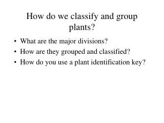
Understanding Living Organisms: Classification and Microscopy Techniques
150 likes | 284 Vues
This article explores how we classify living things based on criteria such as energy needs, environmental responses, reproductive capabilities, and cellular composition. It differentiates between prokaryotic and eukaryotic cells and discusses their structural organization, including organ systems and organelles. Additionally, various microscopy techniques are outlined, including light microscopes (compound and stereo), electron microscopes (SEM and TEM), X-rays, MRI, CT scans, and ultrasound. These tools are vital for studying the intricate structures of life at a cellular level.

Understanding Living Organisms: Classification and Microscopy Techniques
E N D
Presentation Transcript
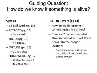
How do we classify something as living? Living things… 1. Have a need for energy (from chemicals, sunlight, other animals, etc.) 2. Respond to their environment (to survive) 3. Have the ability to reproduce= pass genetic info (asexual or sexual) 4. Are made up of one or more cells
StructuralOrganization Animal Large Organ Systems Organs Tissues Cells Organelles Membranes Macromolecules Simple molecules Small Atoms
2 Types of Cells • 1. Prokaryotes= No nucleus (“before” nucleus) • Example = Bacteria
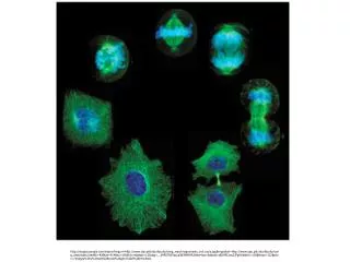
(Kingdom Fungi) • 2. Eukaryotes= Nucleus (“true” nucleus) All other Kingdoms: • Fungi • Plants • Protists • *Animals Eukaryotic cells have many organelles- Structures that perform specific functions. *plasma membrane, cytoplasm, nucleus (Kingdom Plantae) (Kingdom Protista) (Kingdom Animalia)
Tools for Viewing Life • Light Microscope • Compound • Stereo/ Dissecting • Electron Microscope • Scanning (SEM) • Transmission (TEM) • X-rays • Ultrasound • MRI = Magnetic resonance imaging
Light Microscopes Compound= 1 optical system *Magnification = Up to 1500x (SHS=400x) (objective x eyepiece) *Images = Upside down and reversed http://ettc.lrhsd.org/archives/Pictures/138-microscopes-lg.jpg http://www.az-microscope.on.ca/images/Ml2100.jpg
Stereo = 2 separate optical systems (for objects that will not fit on a slide) *Magnification = Up to 100x (SHS = 30x) *Images = 3D, normal upright, right to left image http://www.microscopyu.com/articles/stereomicroscopy/stereointro.html http://www.clt.astate.edu/mhuss/stereoparts.jpg
Electron Microscopes • Scanning (SEM) *Uses electrons instead of light to form/focus image *Used to view surfaces of objects (electrons deflect off specimens) *Magnification up to 500,000x Fly Head http://image53.webshots.com/53/8/76/41/2484876410085329142tuvzFM_fs.jpg http://gsc.nrcan.gc.ca/labs/ebeam/images/sem8.jpg
Transmission (TEM) *Uses electrons instead of light to form/focus image *Used to view inner structure of objects (electrons pass through specimens) *Magnification up to 1, 000,000x TEM-micrograph: thylakoid system in a chloroplast (bar= 0.5 µm). http://www.iopb.res.in/~bhupen/tem_mch.gif http://images.google.com/imgres?imgurl=http://www-classic.uni-graz.at/pphwww/elmi/tem2.jpg&imgrefurl=http://www-classic.uni-graz.at/pphwww/elmi/tempraeparatione.htm&usg=__a3m7XGYsX7CRKvKD0cd0qlD36jk=&h=321&w=387&sz=43&hl=en&start=21&um=1&tbnid=Ugh374nux3hqcM:&tbnh=102&tbnw=123&prev=/images%3Fq%3DTEM%26ndsp%3D18%26hl%3Den%26sa%3DN%26start%3D18%26um%3D1
TEM vs. SEM http://www.vcbio.science.ru.nl/images/TEM-SEM-electron-beam.jpg
X-Ray • X-rays pass through tissue to show dense material (which absorbs the rays) http://www.designswan.com/wp-content/uploads/2008/xray/22.jpg http://www.antonine-education.co.uk/physics_gcse/Unit_1/Topic_5/em_spectrum.jpg
MRI • Uses a magnetic field and radio waves to create detailed images of body tissues. *Can take images from almost every angle http://www.magnet.fsu.edu/education/tutorials/magnetacademy/mri/images/mri-scanner.jpg http://www.ct-scan-info.com/images/MRscanner.jpg
CT-Scan “cat scan” • Computer (Axial) Tomography • Numerous x-ray beams and a set of electronic x-ray detectors rotatearound you, measuring the amount of radiation being absorbed throughout your body • Can create a 3D view of scanned item (which can include items than dense bone) other • Greater number of X-ray images = more radiation http://www.sciencecodex.com/aggregated-images/body/mM1Wb9z7m7ZV3Stj.jpg http://benchmarks.cancer.gov/wp-content/uploads/2010/11/CT-cartoon2.gif
Ultrasound • High-frequency sound waves pass through the body until they come to a border between two tissues that conduct sound differently. Then, some of the sound waves bounce back & are produced as a picture. *When used for long periods of time at high intensities, it can cause the tissues to become heated. http://www.nlm.nih.gov/medlineplus/ency/images/ency/fullsize/18056.jpg http://www.hip2b2.com/images/uploaded_images/Ultrasound.jpg
PET scan • Positron Emission Tomography. • Uses radiation to trace a radioactive element/medicine in the body. • Produces a 3D, color image. http://www.drugs.com/health-guide/images/205902.jpg http://www.petscaninfo.com/zportal/portals/pat/basic/old/pet_scan_cancer.jpg