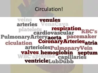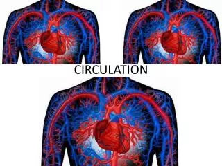
Open circulation
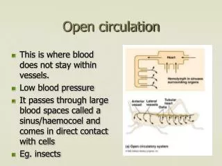
Open circulation. This is where blood does not stay within vessels. Low blood pressure It passes through large blood spaces called a sinus/ haemocoel and comes in direct contact with cells Eg . insects. The Human cardiovascular system. Consists of: Blood Blood vessels Heart

Open circulation
E N D
Presentation Transcript
Open circulation • This is where blood does not stay within vessels. • Low blood pressure • It passes through large blood spaces called a sinus/haemocoel and comes in direct contact with cells • Eg. insects
The Human cardiovascular system • Consists of: • Blood • Blood vessels • Heart • As the blood always remains within vessels it is called a closed circulatory system.It does not come in direct contact with body cells • Advantages: higher blood pressure maintained,faster flow of oxygen & nutrients • Can be more responsive to change and direct blood to where it is needed by constriction & dilation of vessels
Single circulation • Blood pressure reduced after flowing through the tiny capillaries of the gills • Slow flow to the rest of the body • Limits rate of delivery of oxygen and nutrients to tissues • Limits activity levels
Double Circulation • The blood passes through the heart TWICE on each circuit of the body • This means the pressure can be increased again after the lungs to ensure faster delivery of materials to tissues(mammals are active and also have to maintain body temperature) • Body-heart-lung-heart-body. • Circulation to the lungs is the PULMONARY CIRCULATION • Circulation to the body is the SYSTEMIC CIRCULATION at a higher pressure than pulmonary to avoid damaging delicate capillaries
The advantage of double circulation is to maintain higher blood pressure in the circulatory system • In single circulation the blood only passes through the heart ONCE on each circuit • Eg Fish • Disadvantage is lower blood pressure in systemic circulation
Blood Vessels • Arteries(smaller = arterioles)-carry blood away from the heart • Veins(smaller = venules)-carry blood back to the heart • Capillaries-tiny,thin walled vessels connect arteries & veins
Arteries transport blood away from the heart at high pressure to the tissues • The properties of the artery wall adapt it for its function :
Endothelium- the inner lining of the artery made up of squamous epithelial cells. Flat and smooth to minimise friction as blood flows.It is folded and can unfold when the artery stretches • Lumen(blood space)- this is small to maintain high blood pressure • Artery wall – this is thick & strong as it contains the protein collagen to withstand the high blood pressure(120mm Hg or 16 kPa)
It contains elastic fibres(protein elastin)- allows them to give when blood is forced through them- so more in walls nearer the heart. • Then they recoil they give the blood an extra push.This also helps even out blood flow(though you can still feel a pulse) • Both contain smooth muscle tissue,more as you get further from the heart. These muscles can contract,narrowing(constricting) the arterioles and control blood flow. When they relax the arteriole dilates.
Veins – Structure & function • Walls thinner – blood pressure much lower so do not need to be so strong • Thinner layers of collagen,elastic fibres and smooth muscle.They do not need to constrict to control flow. • Larger lumen to ease blood flow. • Valves to prevent backflow
Movement of blood through veins • The low blood pressure means it is harder to return the blood to the heart(less push) • Valves ensure there is no back flow • The valves are semi-lunar or pocket valves
Movement of blood through veins • Many veins run close to skeletal muscles. • Contraction of these muscles squeezes the veins and helps move blood through them
Blood Pressure • The pressure of blood pushing against the walls of the blood vessel. • Mammals have a high blood pressure • This is useful as it maintains a fast flow of blood through the circulatory system.
A cuff that inflates is wrapped around your upper arm and kept in place with Velcro. A tube leads out of the cuff to a rubber bulb. • Air is then blown into the cuff and increasing pressure and tightening is felt on the upper arm. • The doctor puts a stethoscope to your arm and listens to the pulse while the air is slowly let out again. • The systolic pressure(Heart contracting) is measured when the doctor first hears the pulse. • This sound will slowly become more distant and finally disappear. • The diastolic pressure(heart relaxing)is measured from the moment the doctor is unable to hear the sound of the pulse. • The blood pressure is measured in terms of millimetres of mercury (mmHg). Measuring blood pressure
Blood Pressure Changes during circulation • Pressure is highest in the aorta having just been pumped out of the left side of the heart • Pressure varies during a heart beat(systolic) when it is higher and during heart relaxation(diastolic) when it is lower. This change in pressure can be felt as a pulse • As blood moves further from the heart the pressure drops- due to friction (peripheral resistance) with the walls of the vessels. Pulse also disappears due to evening of flow • Largest drop occurs in the arterioles due to increase in area of wall touching the blood.
5. Passing through the narrow capillaries sees a further drop in pressure. Also caused by loss of tissue fluid from the capillaries 6. Venous blood is low in pressure normally about 5 mm Hg 7. Blood returns to the right side of the heart and is pumped to the lungs by the right ventricle.Thinner walls than left side so lower pressure(again surges in the pressure are seen) 8. Again pressure drops while passing through the lung arterioles & capillaries and stays low until returning to the left side of the heart.
Capillary – Structure & Function • Connect arterioles to venules • Take blood as close as possible to all cells in the body – red blood cell may be as little as 1μm from a cell • Allowing rapid transfer of materials between cells & blood • Tiny size allows this- 7μm diameter • Similar size to red blood cells which must squeeze through-slows flow so oxygen can be exchanged presses them close to the capillary wall
Walls of capillaries are only made up of a single layer of endothelial cells – very thin for fast and easy diffusion • Cells have gaps between them making them leaky- tissue fluid seeps out into the cells
Plasma & Tissue Fluid • The liquid part of the blood is plasma it carries the blood cells and many dissolved materials
Tissue Fluid • As blood flows through the capillaries fluid leaks out into the spaces around your cells(one sixth of your body!!) • This is TISSUE FLUID-it surrounds our cells and allows exchange of materials-will contain O2,nutrients etc • Identical to plasma except does not contain red blood cells,though some white cells can squeeze out • Contains less proteins as these are too large to pass out
Factors controlling movement of tissue fluid • Blood pressure: (hydrostatic pressure) forces fluid out at the arteriole end of the capillary bed • Osmotic pressure (solute pressure) : returns fluid to the blood at the venule end • Overall net flow OUT into the tissues
Arterial end • Effective HP = 4.3-1.1 = 3.2kPa(out) • Effective SP = -3.3-(-1.3) = -2kPa(in) • Overall effective blood P = 3.2-2=1.2 kPa • Fluid pushed out • Venule end • Effective HP = 1.6-1.1=0.5 kPa(out) • Effective SP =-3.3-(-1.3)=-2kPa(in) • Overall effective blood P= 0.5 -2 = -1.5 kPa • Fluid pushed back into capillary Arteriole end Venule end 4.3 -3.3 1.6 -3.3 1.1 -1.3 1.1 -1.3 Net outflow 1.2kPa Net inflow -1.5kPa Hydrostatic pressure solute pressure (water potential)
The Lymphatic system • About 90% of fluid that leaks from the capillaries returns to them • 10% does not - and must be drained away by lymph vessels • These vessels form the lymphatic system
Lymph Vessels • Tiny,dead end vessels which are found in almost all tissues • Fluid enters them through tiny valves • This fluid is called lymph and is virtually identical to tissue fluid
Lymph vessels • The tiny valves allow large proteins to enter the lymph(cannot enter the blood capillary) • With out them protein would build up in your tissue fluid causing oedema • (Water leaving your blood causing swelling of tissues) • And death within 24 hours
Lymph • In some parts of the body the lymph has differences: • Small intestine lymph will contain high amounts of lipids after a meal • The villi contain a lymph vessel(a lacteal),which absorbs digested fats
Lymph • Tissue fluid and lymph in the liver has high concentrations of proteins. • Lymph is moved through the vessels very slowly(100cm³/h – blood 80cm³/sec!) • Contraction of skeletal muscles help this and • also the vessels have smooth muscle in their walls which contracts to squeeze lymph along • Lymph eventually drains back into the blood in the subclavian veins
Lymph Nodes • Found at intervals in the lymphatic system • Involved in protection from disease • Microbes are filtered from the lymph as it passes through the lymph nodes by white blood cells - lymphocytes
The cardiac cycle • The heart beats on average 70bpm • The cardiac cycle is the events that take place in one heart beat.
The cardiac cycle • Atrial systole(0.1s) Heart full of blood . Atria walls contract, blood pushed through the AV valves into the ventricles.Valves in the vena cava and pulmonary vein prevent backflow.
2. Ventricular systole(0.3s) Thick ventricle muscle walls contract.This produces a higher pressure.Blood is forced into the PA and aorta, through the semilunar valves which are forced open. • Papillary muscles also contract to pull on the tendons, stopping the closed AV valve flaps turning inside out.
3.Ventricular diastole Ventricle and atrial muscles are relaxed. heart begins to fill with blood starting in the atria . Blood pressure in the aorta and pulmonary artery shuts the semi lunar valves
Heart valves • Semilunar valve • Consists of 3 pockets in half moon shape • Blood flowing through pushes past the pockets • When it tries to return it will fill the pockets closing the valve.
Pressure changes in the heart during the cardiac cycle (a) Atrial systole (b) ventricular systole • Atria contract (atrial systole) • ventricular systole begins. When pressure in the ventricle exceeds pressure in the atria AV valves shut. • Ventricular systole,rapid increase in pressure as ventricles contract • Pressure in ventricle exceeds that in aorta, semilunar valves open. • Maximum systolic pressure,ventricles now start to relax • Pressure in ventricles drops below aorta.SL valves shut • Wave of contraction running down aorta raises pressure • Ventricular diastole starts • Blood starts to refill the atria increasing pressure • When pressure exceeds ventricle, AV valves open and blood flows into the ventricles