Download
1 / 26
280 likes | 343 Vues
Muscles of the Anterior Neck and Throat: Suprahyoid. Four deep throat muscles Form the floor of the oral cavity Anchor the tongue Elevate the hyoid Move the larynx superiorly during swallowing. Muscles of the Anterior Neck and Throat: Suprahyoid. Figure 10.8a.

E N D
Muscles of the Anterior Neck and Throat: Suprahyoid • Four deep throat muscles • Form the floor of the oral cavity • Anchor the tongue • Elevate the hyoid • Move the larynx superiorly during swallowing
Muscles of the Anterior Neck and Throat: Suprahyoid Figure 10.8a
Muscles of the Anterior Neck and Throat: Infrahyoid • Straplike muscles that depress the hyoid and larynx during swallowing and speaking
Muscles of the Anterior Neck and Throat: Infrahyoid Figure 10.8b
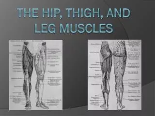
Muscles of the Neck: Head Movements • Major head flexor is the sternocleidomastoid • Synergists to head flexion are the suprahyoid and infrahyoid • Lateral head movements are accomplished by the sternocleidomastoid and scalene muscles • Head extension is accomplished by the deep splenius muscles and aided by the superficial trapezius
Muscles of the Neck: Head Movements Figure 10.9a
Muscles of the Neck: Head Movements Figure 10.9b
Trunk Movements: Deep Back Muscles • The prime mover of back extension is the erector spinae • Erector spinae, or sacrospinalis, muscles consist of three columns on each side of the vertebrae – iliocostalis, longissimus, and spinalis • Lateral bending of the back is accomplished by unilateral contraction of these muscles • Other deep back extensors include the semispinalis muscles and the quadratus lumborum
Trunk Movements: Deep Back Muscles Figure 10.9d
Trunk Movements: Short Muscles • Four short muscles extend from one vertebra to another • These muscles are synergists in extension and rotation of the spine Figure 10.9c
Muscles of Respiration: External Intercostals • The primary function of deep thoracic muscles is to promote movement for breathing • External intercostals – more superficial layer that lifts the rib cage and increases thoracic volume to allow inspiration Figure 10.10a
Muscles of Respiration: Internal Intercostals • Internal intercostals – deeper layer that aids in forced expiration • Diaphragm – most important muscle in inspiration Figure 10.10a
Muscles of Respiration: The Diaphragm Figure 10.10b
Muscles of the Abdominal Wall • The abdominal wall is composed of four paired muscles (internal and external obliques, transversus abdominis, and rectus abdominis), their fasciae, and their aponeuroses • Fascicles of these muscles run at right and oblique angles to one another, giving the abdominal wall added strength
Muscles of the Abdominal Wall • In addition to forming the abdominal wall, these muscles: • Are involved with lateral flexion and rotation of the trunk • Help promote urination, defecation, childbirth, vomiting, coughing, and screaming
Muscles of the Abdominal Wall Figure 10.11a
Muscles of the Abdominal Wall Figure 10.11b
Muscles of the Abdominal Wall Figure 10.11c
Muscles of the Pelvic Floor (Pelvic Diaphragm) • The pelvic diaphragm is composed of two paired muscles – levator ani and coccygeus • These muscles: • Close the inferior outlet of the pelvis • Support the pelvic floor • Elevate the pelvic floor to help release feces • Resist increased intra-abdominal pressure
Muscles of the Pelvic Floor: Pelvic Diaphragm Figure 10.12a
Muscles Inferior to the Pelvic Floor • Two sphincter muscles allow voluntary control of urination (sphincter urethrae) and defecation (external anal sphincter) • The ischiocavernosus and bulbospongiosus assist in erection of the penis and clitoris
Muscles of the Pelvic Floor Figure 10.12b
Muscles of the Pelvic Floor Figure 10.12c
Extrinsic Shoulder Muscles • Muscles of the thorax • Anterior: pectoralis major, pectoralis minor, serratus anterior, and subclavius • Posterior: latissimus dorsi, trapezius muscles, levator scapulae, and rhomboids • These muscles are involved with the movements of the scapula including elevation, depression, rotation, and lateral and medial movements • Prime movers of shoulder elevation are the trapezius and levator scapulae
Extrinsic Shoulder Muscles Figure 10.13a
Extrinsic Shoulder Muscles Figure 10.13b