Insect Transmitted Nematodes
Insect Transmitted Nematodes. Filarial worms Tissue dwelling parasites Order Spirurida Transmitted to definitive host through insect bite. Filarial Nematodes . Tissue-dwelling nematodes (not in digestive tract) Possess a unique life cycle stage – the microfilaria - between the egg and J 1


Insect Transmitted Nematodes
E N D
Presentation Transcript
Insect Transmitted Nematodes • Filarial worms • Tissue dwelling parasites • Order Spirurida • Transmitted to definitive host through insect bite
Filarial Nematodes • Tissue-dwelling nematodes (not in digestive tract) • Possess a unique life cycle stage – the microfilaria - between the egg and J1 • Egg microfilaria J1 J2 J3 J4 Adult • these are present in the bloodstream or skin of the definitive host. • Filarial worms utilize arthropods as vectors.
Lymphatic Filariasis • Wucheria bancrofti • Brugia malayi • Lymphatic filariasis • 119 million infected • Elephantiasis • Manifestation of lymphatic filariasis
Distribution of Wucheria bancrofti • Broad equatorial belt • Africa, Middle East, Southeast Asia, Indo-Pacific islands, Parts of Australia and South America
Habitat • Adults live in lymphatic ducts. • Usually near major lymph glands in lower half of body • Release juveniles (microfilariae) into lymph • Microfilariae carried to blood stream
Lymphatic System • Network of vessels that collect fluid that leaks out of the blood into tissues (lymph) • Redirects lymph back into the blood stream
Adults of Wuchereria bancrofti Adults occur in the lymphatic vessels
Wuchereria bancrofti • Females release juveniles into lymph (ovoviviparous) • microfilariae swept into blood stream • Mosquitoes ingest microfilariae with blood meal
Wuchereria bancrofti • Penetrate stomach of mosquito • Develop in thoracic muscles • Develop into filariform juveniles
Wuchereria bancrofti • Migrate to the proboscis • Injected into human with blood meal • Mature in lymphatic ducts
Periodicity • Microfilariae in peripheral blood at periodic intervals • Wucheria bancrofti • In peripheral blood between 10:00pm-2:00am • In blood of deep tissues during the day • Coincides with feeding time of intermediate hosts
Microfilariae of Wuchereria bancrofti Nocturnal periodicity of microfilariae
Pathogenesis • Depends on inflammatory and immune response • Clinical manifestation varies
Phases of Pathogenesis • Asymptomatic Phase High levels of microfilaremia Immune response down regulated Sometimes no symptoms and no microfilaremia • People in endemic areas Sometimes lymphatic inflammation and no microfilaremia • Travelers who get infected
Phases of Pathogenesis 2. Inflammatory (Acute) Phase Caused by antigens from adult worms Inflammation due to bacterial infection Adults interfere with lymph flow • Lymphedema • Inflammation of lymph channels • Inflammation of lymph nodes • Symptoms: • Chills • Fever • Swollen and painful lymph nodes • Swelling of reproductive organs • Lasts 5-7 days
Phases of Pathogenesis 3. Obstructive (Chronic) Phase • Lymph ducts become blocked • Fibrosis of infected areas • Swelling • Accumulation of lymph • Elephantiasis: accumulation of lymph in extremeties, fibrosis, and thickening of skin. • Chyluria (lymph in the urine)
Affected Areas • Legs • Scrotum • Arms • Brest
Pathology of Wuchereria bancrofti Obstructive phase photos
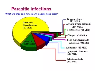
Impacts • Rarely fatal • Disfiguring • 40 million people • Disability • Daily functions • Sexual disability • WHO: second leading cause of permanent and long-term disability in the world (after leprosy) • Social impacts
Diagnosis • Demonstration of microfilariae in blood • PCR diagnosis
Microfilariae of Wuchereria bancrofti • Microfilariae are seen in blood smears and are DIAGNOSTIC • worms are 230-320 µm long
Treatment • Diethyl-carbamazine and Ivermectin • Kills adults and microfilariae • Edematous limbs • Pressure bandages • Surgical removal of elephantoid tissue
Epidemiology • Timing • Takes 6-12 months for females to release microfilariae • Produce microfilariae for 5-10 years • How do you get infected? • Bite from infected mosquito
Brugia malayi Causes Malayan filariasis Distribution - Orient, South Pacific, and Southern Asia to India – overlaps with Wuchereria bancrofti - but does not occur in Africa or South America
Brugia malayi Morphology and life cycle is similar to that of Wuchereria bancrofti
Brugia malayi Pathology - Adults live in lymphatic vessels of the arms and legs and cause elephantiasis in these regions Difference from Wuchereria?
Stages of Dirofilaria immitis Adult male: 6-12 inches long Adult female: 12-16 inches long Adults coiled in right side of dog heart Unsheathed microfilaria in dog blood - DIAGNOSTIC
Pathology of Dirofilaria immitis • PATHOLOGY caused by adult worms. • First signs of infection involve exercise intolerance • due to inadequate blood supply to lungs • infected dogs cough, have shortness of breath, and tire rapidly. • 2. Eventually the dog suffers congestive heart failure- usually after a period of exercise.
Dirofilaria immitis • PREVENTION - chemoprophylaxis • 2 drugs are used: ivermectin (in Heartgard) and milbemycin oxime (in Sentinel and Interceptor) • - How taken? • How does it work? • How long to treat?
Human Cases of Dirofilaria immitis • HUMAN INFECTIONS of Dirofilaria immitis are rare (~70 cases). • Larvae are killed by the host reaction and scar tissue nodules form in lungs around worms • Symptoms are coughing and chest pain. • In only 4 cases were adult worms recovered from the human heart. These were found incidentally at autopsy and were not related to the death of the patient.
Onchocerca volvulus Causative agent of Onchocerciasis or River Blindness DISTRIBUTION – Areas of Africa, Arabia, Guatemala, Mexico, Venezuela and Colombia
Life Cycle of Onchocerca volvulus • Nodules are most common below the waist in region of Africa. • Nodules are on the head and above the waist in Central & South America.
Life Cycle of Onchocerca volvulus 1. Adults live in coiled masses encapsulated under the skin.
Life Cycle of Onchocerca volvulus 2. Females produce microfilariae - Microfilariae of Onchocerca NEVER invade the bloodstream.
Life Cycle of Onchocerca volvulus 3. Microfilariae in the skin are ingested by the black fly intermediate host, Simulium damnosum, when a blood meal is taken.
Life Cycle of Onchocerca volvulus 4. Parasites develop to J3’s in the musculature of the black fly and migrate to the mouthparts. 5. J3’s are inoculated into the skin when black fly bites. Adults mature in a year within subcutaneous nodules.
Adults of Onchocerca volvulus Microscopic section showing adults and scar tissue reaction around them forming the nodule Skin nodule cut open to reveal adults coiled together
Microfilariae of Onchocerca volvulus Unsheathed microfilariae occur in the skin, never the bloodsteam
Pathology of Onchocerca volvulus ADULTS cause onchocercomas Nodules are about ½ -1 inch in diameter. Nodules are relatively benign and cause only some disfigurement.
Pathology of Onchocerca volvulus • MICROFILARIAE cause 3 severe problems. This is the only filarial worm in which microfilariae are pathogenic! • 1. Microfilariae in the skin cause severe dermatitis • skin becomes thickening, • discoloration, and cracking. • leading to secondary bacterial infections • - itching is so severe some people have committed suicide
Pathology of Onchocerca volvulus 2. Microfilariae in skin cont: - in parts of Africa, the skin of the scrotum and inguinal area loses its elasticity causing hanging groin!