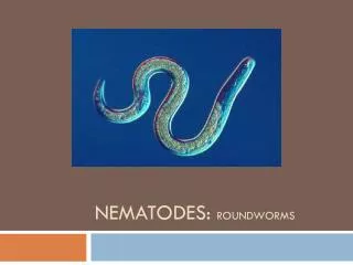
Intestinal Nematodes
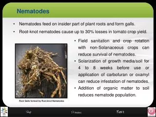
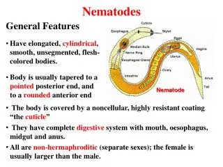
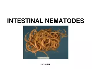
Intestinal Nematodes . Classification of Parasites. Elongated worm, cylindrical, unsegmented and tapering at both ends. Variable in size, measure <1 cm to about 100cm. Sex separate and male is smaller than female. Nematodes : General features. Nematodes: Location in the human body .

Intestinal Nematodes
E N D
Presentation Transcript
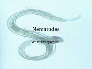
Elongated worm, cylindrical, unsegmented and tapering at both ends. Variable in size, measure <1 cm to about 100cm. Sex separate and male is smaller than female Nematodes :General features
Nematodes: Location in the human body • Intestinal nematodes • Tissue nematodes
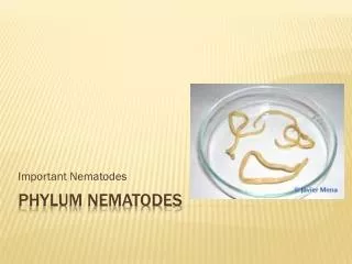
Common intestinal nematode infections: Enterobius (Oxyuris) vermicularis(Pinworm,seatworm,threadworm) Trichuristrichiura(whipworm) Ascarislumbricoides(roundworm) Ancylostomaduodenale & Necatoramericanus(hookworms) Strongyloidesstercoralis : Nematodes: common intestinal infections
(Common names :Pin worm, seat worm, thread worm( Found all over the world. adult in lumen of cecum and appendix from which adult female migrate to rectum. It can be seen by naked eye as white thread ± 1cm. Male is smaller than female ± 0.5cm, with coiled end. Enterobiusvermicularis (Oxyuris)
Enterobiusvermicularis (Oxyuris) LIFE CYCLE
Pathology Majority of infections are asymptomatic. Main clinical presentation pruritusani perianal excoriation Ectopic enterobiasis occurs in female when invade vulva and vagina result in valvovagintis Usually accompanied by insomnia, anorexia, loss of weight and concentration (Side effect) Enterobiusvermicularis (Oxyuris)
Treatment ِِAlbandazole , Mebendazole for whole family Enterobiusvermicularis (Oxyuris)
Ascarislumbricoides (roundworm) Ascaris adult
The commonest human helminthes infection. Found in jejunumand upper part of ileum. Female ± 20 cm longer than male ± 10 cm Feed on semi digested food. Ascarislumbricoides (roundworm)
Ascarislumbricoides (roundworm) LIFE CYCLE
Ascarislumbricoides (roundworm) Ascaris egg (embryonated)
Ascaris eggs Ascaris larva emerging from egg Ascaris egg (embryonated)
Pathology: 1-Adult worm: Light infection : asymptomatic. Heavy infection : intestinal obstruction Migrating adult : to bile duct -jaundice 2-Larvae: Loeffler`s syndrome (imp) Pneumonia, cough with bloody sputum Eosinophilia, urticaria Ascarislumbricoides (roundworm)
Ascarislumbricoides (roundworm) Loeffler`s syndrome:Larvae in lung pnumonia,cough ,bloody sputum
Ascarislumbricoides (roundworm) Ascaris larva in lung
Diagnosis: -eggs in stool. -larvae in sputum. -adult may pass with stool. Ascarislumbricoides (roundworm) Treatment: Albendazole , Mebendazole
Trichuris trichiura (Whipworm) LIFE CYCLE
World wide ,common in poor sanitation. It coexists with Ascaris because of similar requirement. Adult live in large intestine especially caecum and appendix–inheavy infection the whole length of large intestine affected. Male and female worm have narrowanterior portion penetrate the intestinal mucosa Trichuristrichiura(whipworm)
Pathology light infection : asymptomatic heavy infection :abdominal pain ,bloody diarrhea. Rectal prolapse in children is a common complication. - Trichuristrichiura(Whipworm)
Trichuristrichiura(Whipworm) Embryonated egg Unembryonated egg Infective stageDiagnostic stage
-Diagnosis: egg in stool characterized by its barrel shape with mucoid plugs at each pole . Treatment :Albendazole. Trichuristrichiura(Whipworm)
Hook worms Ancylostomadudenale &Necatoramericanus
Hook worms Buccal cavity attached to intestinal mucosa
Hook worms Ancylostomadudenale &Necatoramericanus LIFE CYCLE
A common cause of anemia. Found in small intestine mainly jejunum. Its buccal capsule (mouth) lined with hard hooks, triangular cutting plates and anticoagulant glands. Hook worms Ancylostomadudenale &Necatoramericanus
Hook worms Pathology& clinical picture: • - larvae: • At the site of entry of larvae (ground itch). • Migration phase: • cough with bloody sputum • pneumonia, eosinophilia,urticaria. • - adult worm: • low worm burden: no symptoms. • Moderate to heavy burden: • Epigastric pain, vomiting , hemorrhagic enteritis. • Protein loss: hypoproteinaemia edema. • Anemia: due to withdrawal of blood by parasites and hemorrhage from punctured sites lead to sever anemia = microcytichypochromic .
Diagnosis: -Eggs in stools.; -occult blood (+) Hook worms Diagnosis and treatment Treatment: Albendazol, Mebendazole
Strongyloidesstercoralis Widely distributed in tropical region worldwide . fetal opportunistic in immuno-compromised host. It is smallest pathogenic nematodes ± 2.5mm. adult live in mucous membrane of duodenum jejunum rarely m.m.of bronchus.
Strongyloides stercoralis LIFE CYCLE
Cuteneouslittle reaction on penetration. sever dermatitis at perianal region in case of external autoinfection. Migration :same as hook worms . Intestinal: inflammation of upper intestinal mucosa, diarrhea, upper abdominal pain clocky in nature. Disseminated strongyloidiasis : in patient with immunodeficiency ,uncontrolled diarrhea –granulomatus changes –necrosis--perforation--peritonitis--death. Strongyloidesstercoralis: Pathology and clinical picture:
Diagnosis: rhabditiform larvae diagnostic stage in: -Stool examination -Duodenal aspirate Treatment:Albandazole, Mebendazole Strongyloidesstercoralis