Download
1 / 18
190 likes | 676 Vues
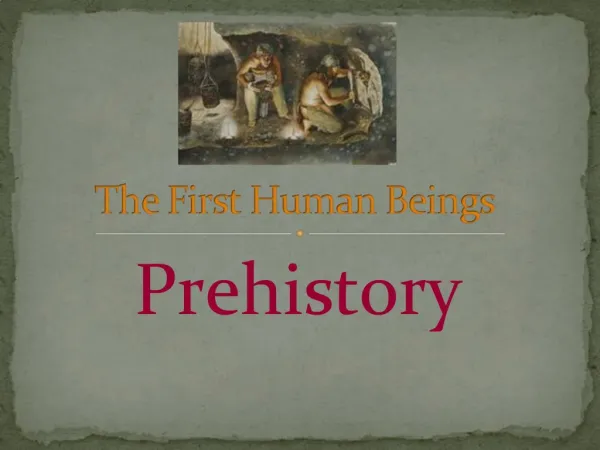
Intestinal Nematodes of Human Beings. Trichinella spiralis. Diseases -Trichinosis, Trichiniasis, Trichinelliasis Morphology -the infrequently seen adult is a small worm. It is characterized by: Slender anterior end with a small, orbicular, nonpapillated mouth

E N D
Trichinella spiralis • Diseases -Trichinosis, Trichiniasis, Trichinelliasis • Morphology -the infrequently seen adult is a small worm. It is characterized by: • Slender anterior end with a small, orbicular, nonpapillated mouth • Posterior end bluntly rounded (female) ventrally curved with two lobular caudal appendages (male) • Single ovary with vulva in the anterior fifth (female) • Long narrow digestive tract
Tail of male Trichinella spiralis • The adult worms measure 1.5 mm (male) to 3.5 mm (female) long
LIFE CYCLE • Animals are infected with Trichinella spiralis when they ingest infective larvae in raw or undercooked meat. The larvae mature into adults in the host's small intestine and the female worms give birth to larvae. (The males die after fertilizing the females, and the females die after producing larvae.) The larvae enter the blood stream of the host and, eventually, end up in the host's muscles. Here the larvae mature into infective larvae, and the next host is infected when it eats these larvae. In the muscles the larvae cause a severe host reaction that results in soreness and tenderness of the muscles. Although this parasite probably only rarely causes fatalities in humans, it can cause extreme discomfort.
EPIDEMIOLOGY • Infected persons in the world has been greatly reduced. The prevalence of Trichinosis is less in the tropics and subtropics, chiefly owing to low consumption of pork products, religious bans on eating pork, Chinese culinary custom of cooking it thoroughly.
People may also acquire the infection by the ingestion of ground beef. Beef ground in a machine in which infected pork has been ground is a possible source of infection. Through an aggressive program of meat inspection, the incidence of trichinosis in pigs in the United States has been lowered to less than 1%, so it is unlikely (but not impossible) that pork products purchased in your local supermarket will contain Trichinella larvae. Most recent outbreaks of trichinosis in the United States have been traced to pork products from pigs that have not been inspected and that have been slaughtered privately. Because of its low host-specificity, almost any "wild" meat should be considered suspect, and hunters should be careful when preparing meat from their kills. In particular, a number of infections have been traced to contaminated bear meat.
PATHOLOGY • The pathology of Trichinosis is concerned with the presence of larvae in the striated muscles and vital organs and with the reaction of the host to their activities. The muscle fiber increased in size, become edematous, develop a spindle shape and undergo basophilic degeneration
Common Diagnostic Test Muscle biopsy, Antibody ELISA • Clinical Signs: Usually asymptomatic. Heavy infections have been reported to cause a hemorrhagic enteritis, muscle pain and stiffness.
Diagnostic Stage: First stage-larva (arrows) in the nurse cell. Larval cysts can get up to 3 mm in diameter.
TRICHURIS TRICHIURA • Diseases –Trichuriasis, trichocephaliasis, whipworm infection. • LIFE CYCLE The adults live in the host's large intestine with their anterior ends embedded in the cells that line the intestine; each female can produce in excess of 10,000 eggs each day, and the worms can live several years. The eggs are passed in the host's feces, and they become infective in about three weeks. When an infective egg is eaten by the appropriate host it hatches in the small intestine, and the worm migrates to the large intestine where it reaches sexual maturity. Most infections of whipworms are probably asymptomatic. However, because the worms live a long time and a person can be reinfected constantly, heavy worm burdens can develop.
Trichuris trichiura adults. Trichinella spiralis in tissue
Trichuris vulpis egg. Trichuris trichiura in the large intestine.
The posterior end of a male Trichuris sp., with an everted spicule