Download

1 / 42
420 likes | 628 Vues
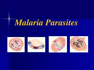
~* MALARIA PARASITES OF HUMAN BEINGS *~. ~* GENUS *~ The malarial parasites of humans are caused by species of the genus Plasmodium of the class Sporozoa . The asexual cycle (schizogony) takes place in the red blood cells of vertebrates and the sexual cycle (sporogony) in mosquitoes.

E N D
~* GENUS *~ • The malarial parasitesof humans are caused by species of the genus Plasmodium of the class Sporozoa. • The asexual cycle (schizogony) takes place in the red blood cells of vertebrates and the sexual cycle (sporogony) in mosquitoes. • The members of this genus, which cause malaria in mammals and birds, have closely similar morphology and life cycles.
~* DISEASES *~ • Malaria, are known by several names among which are paludism, intermittent fever, chills and fever, Roman fever, Chagres fever, marsh fever, tropical fever, coastal fever and ague. • The term malaria is derived from two Italian words, mal (bad) and aria (air).
~* Species Pathogenic to Man *~ • P. malariae was described in 1880 by Laveran. • ~ has an affinity for mature or older red cells • P. vivax was named in 1890 by Grassi and Feletti. • ~ prefer to invade young red cells • P. falciparum in 1897 by Welch. • ~ infects cells of all ages • P. ovale in 1922 by Stephens. • ~ prefer to invade young red cells
~* MORPHOLOGY *~ • Some general characteristics are common to all malaria parasites, but differential features make it possible to identify species. • The earliest form after invasion of the re blood cell is a ring of bluish cytoplasm with a dot-like nucleus of red chromatin. • As this early stage, the trophozoite, grows the erythrocyte hemoglobin is metabolized to produce a darkly staining malarial pigment, hemozoin.
Depending upon the speciesof parasite, the cytoplasm may become irregular in shape, and the red blood cell may show pink granules. • When the growing parasite divide, it is called a schizont, showing multiple masses of nuclear chromatin. • Some of the trophozoites develop into gametocytes, or sexual stages, which are differentiated by compact cytoplasm and the absence of nuclear division.
~* PLASMODIUM VIVAX *~ • The infected red cell is enlarged, but this may be partly explained by the affinity of the parasite for larger reticulocytes. • As the signet-ring appearing trophozoite grows, it becomes irregular in shape, with amoeboid extensions of the cytoplasm. • Schuffner’s dots make their appearance in properly stained smears of P. vivax infected cells; these are fine, round, pink or reddish granules, distributed uniformly over the red cell. • Increasing amounts of the of pigment accumulate in the parasite cytoplasm.
After 36 hours, the parasite fills over half the enlarged red cell, and the nucleus divides, becoming a schizont. • By 48 hours the schizont has segmented into 16 distinct cells, the merozoites, each with a red nucleus and blue cytoplasm condensed about it. And the rupture of the erythrocyte takes place. • The gametocytes resemble a late trophozoite prior to segmentation. • They are oval, nearly filling the red cell – the microgametocyte with less deeply staining nucleus and cytoplasm, the macrogametocyte with darker blue cytoplasm and more compact nucleus.
RING RING RING RING TROPH TROPH TROPH TROPH AMOEBOID SCHIZONT SCHIZONT SCHIZONT
SCHIZONT IMMATURE SCHIZONT MATURE SCHIZONT GAMETOCYTE GAMETOCYTE GAMETOCYTE FEMALE GAMETOCYTE MALE GAMETOCYTE
~* PLASMODIUM MALARIAE *~ • The early ring form of P. malariae resembles that of P.vivax, but the parasite is smaller, less irregular, and more compact, the cytoplasm is a deeper blue. • The growing trophozite acquires coarse granules of dark brown or black pigment and may assume a band shape across the cell. • Infected red cells are normal or even smaller than most in size since old cells are preferentially infected. • A period of 72 hours is required for the development of the mature schizont, which resembles a daisy or rosette with only eight to ten oval merozoites.
A compact mass of greenish black pigment is often located centrally, surrounded by merozoites. • The gametocytes are similar to those of P. vivax but are smaller.
RING TROPH TROPH COMPACT TROPH BAND FORM IMMATURE SCHIZONT MATURE SCHIZONT SCHIZONT SCHIZONT SCHIZONT SCHIZONT FEMALE GAMETOCYTE
GAMETOCYTE GAMETOCYTE
~* PLASMODIUM FALCIPARUM *~ • This differs from other plasmodia of humans in that, except in infections with very high parasitemia, only in the ring forms of early trophozoites and the gametocytes are ordinarily seen in the peripheral blood. • Schizogony takes place in the capillaries of the muscles and viscera, and very few schizont are found in the peripheral blood. • The infected red blood cells are of normal size. • The presence of more than one ring form in a cell is relatively common.
Double chromatin dots are frequently found in P. falciparum ring forms, and only occasionally in the rings of other species. • The schizonts, seldom found in the blood, resemble those of P. vivax but have smaller and a few more merozoites when mature and ready to rupture. • The immature gametocytes gradually acquire an elliptical shape, stretching but remaining within the red cell. • When fully developed, they have a characteristic banana shape, the so-called crescent. • In cells, infected with P. falciparum there are sometimes cytoplasmic precipitates known as Maurer’s dots that appear as irregularly distributed red spots or clefts.
APPLIQUE FORM DOUBLE RING MULTIPLE RING OLD RING RING TROPHOZOITES MATURE SCHIZONT GAMETOCYTE MALE GAMETOCYTES
~* PLASMODIUM OVALE *~ • Commonly found in West Africa, but infrequently encountered elsewhere, is similar to P. vivax and P. malariae in several characteristics. • The infected red blood cells are slightly enlarged, can be of oval shape, and show Schuffner’s dots. • An important diagnostic feature is the irregular or fimbriated appearance of the edges of the infected red cell. • Schizonts have centrally massed pigment and only about eight merozoites when mature.
RING RING RING TROPH TROPH TROPH SCHIZONT SCHIZONT MATURE SCHIZONT
~* LIFE CYCLE *~ • The life of the plasmodia is passed in two hosts, a vertebrate and a mosquito. • The asexual cycle in the vertebrate host is known as schizogony, and the sporulating sexual cycle in the mosquito as sporogony.
~* SCHIZOGONY *~ • The infectious sporozoite from the salivary glands of an infected female Anopheles mosquito is injected during biting into the human blood stream. • Within 30 minutes this slender motile organism enters a liver parenchymal cell, initiating what is called the exoerythrocytic (EE) potion of the life cycle, because the red blood cells have not yet been invaded. • Within the liver cell, the parasite begins an extensive multiplication and is called a schizont, producing thousands of merozoites within the liver cell after 8 to 15 days, depending upon the species of malaria.
The parasitized liver cell then ruptures, freeing merozoites to initiate the erythrocytic cycle. • A relapse signifies that parasitemia develops from EE stages in the liver. • A recrudescence means an increase in parasites that has persisted at low levels in the blood. • The erythrocytic cycle consists of the invasion of red cells by merozoites, their development through trophozoites, then schizonts, the rupturing of the cell, and the reinvasion of new cells. • The process of invasion involves recognition by the merozoite of a specific receptor site on a red cell membrane and proper orientation of the anterior end of the merozoite, exposing special organelles to the red cell surface.
Then, after a few seconds, the red cell becomes deformed and the merozoite enters through a invagination of the red cell membrane. • The red cell for P. vivax malaria has been found to be associated with the Duffy blood-group antigen. • As repeated cycles of asexual multiplication occur, some parasites that invade the red cells do not undergo division as schizonts but, instead, transform into male and female gametocytes.
~* SPOROGONY *~ • Sporogony, the sexual cycle, takes place in the mosquito. • The gametocytes ingested with the blood meal, unlike the schizonts, are not digested. • In the male microgametocyte, the chromatin dot divides into 6 to 8 nuclei that migrate to the periphery of the parasite. • There, several whip-like, actively motile filaments, the unicellular microgametes, are thrust out and detached from the parent cell, a process known as exflagellation. • In the meantime, the female macrogametocyte has matured into a macrogamete.
Fertilization is achieved by the entry of the microgamete, forming a zygote. • In 12 to 24 hours after the mosquito’s blood meal, the zygote changes into a worm-like form, the ookinete, which penetrates the wall of the mosquito’s gut and develops into a spherical oocyst between the epithelium and the basement membrane. • It increases to many times its original size, with thousands of sporozoites developing inside. • With rupture of the oocyst, the sporozoites are liberated into the body cavity and migrate to the salivary glands. • When the mosquito feeds on humans, the sporozoites gain access to the blood and tissues and enter upon their exoerythrocytic cycle.
The sporogonic cycle in the mosquito takes from 10 – 17 days. • Transmission of malaria occurs by other mechanisms, such as blood transfusion, contaminated syringes, or across the placenta. • Malaria may be accidentally transfused from an infected but asymptomatic donor to a recipient. • Malaria outbreaks have resulted from multiple use of a syringe among “mainlining” drug addicts. • Transfusion of malaria initiates only the erythrocytic cycle and therefore is usually eradicated, since there is no exoerythrocytic cycle. • A rare type of non-mosquito transmission is congenital infection. When it does occur it indicates some placental defect, and it can involve any species of parasite.
~* PATHOLOGY *~ • The pathologic changes associated with all types of malaria have certain features in common, but the best known are those of P. falciparum malaria, the cause of virtually all fatalities. • The reticuloendothelial system is activated by the rupture of infected red cells and intravascular releaseof parasites, malarial pigment, and cellular debris. • The liver and spleen bear the brunt of disposing of this particulate material by hyperplasia of Kupffer cells and macrophages, resulting in enlargement of the liver and spleen.
In acute infections the soft, enlarged spleen is susceptible to spontaneous or traumatic rupture. • Fixed macrophages of the spleen and liver exhibit phagocytosis of infected and even some normal erythrocytes, as well as the presence of ingested malarial pigment. • With the increased red cell destruction there is increased turn over of iron and blood pigments, and excess oron not immediately used to form new hemogoblin is deposited as hemosiderin in parenchymatous cells. • In chronic or repeated infections the liver and spleen especially, but also other organs, become slate-gray or black in color from the deposition of malarial pigment. • Continued activity of malaria leads to a firm, fibrotic spleen that shrinks back to normal size.
Hepatic dysfunction is minimal, showing only slight elevation of bilirubin and liver enzyme levewls. There is little or no reaction to the exoerythrocytic cells. • If the malarial infection continues, anemia develops, and often the anemia is of greater degree than can be explained by direct destruction of red cells by the parasite. • The complement system is activated, with consumption of complement components via the classical pathway. • Immune complexes are formed and may be deposited in the kidneys. • Blackwater fever is a term given to the clinical syndrome of acute and massive hemolysis during malaria.
~* SYMPTOMATOLOGY *~ • The incubation period which generally varies from 9 – 30 days, may be prolonged for months. • Small numbers of parasites are actually present in the circulation a day or two before the symptoms begin. • As the numbers of parasites increase, the infected individual begins to experience various general symptoms, such as headache, lassitude, vague pains in the bones and joints, chilly sensations, and fever. • Within a few more days regular episodes of chills and fever become more prominent, but other systemic symptoms, especially headache, muscle aches, and pains, persist.
The malarial paroxysm occurs at the end of the schizigonic cycle, when the merozoites of the mature schizonts, together with their pigments and residual erythrocyte debris, erupt from infected red cells and are released into the circulation. • The malarial paroxysm is one of the most dramatic and frightening events in clinical medicine. It begins with a cold stage up to an hour, with chilly sensations that progress to teeth-chattering, frankly shaking chill. Peripheral blood vessels are constricted and the lips and nails are cyanotic. • At the end of the cold stage the body temperature begins to mount rapidly, ushering in the hot stage, which lasts from 6 to 12 hours.
Nausea and even vomiting, headache, and a rapid pulse occur; the circulation opens up, and the temperature peaks at 103 to 106 F (39 to 41 C), with the skin hot and the face flushed. • The patient may be euphoric; the high fever may produce convulsions in children. Then the patient perspires profusely; the temperature falls and the headache disappears. • In a few hours, the patient is exhausted but symptomless. The next day the patient can feel quite well before the next paroxysm occurs. • When malarial paroxysms are discrete and occur at regular intervals, they are said to be synchronized. • Synchrony depends upon all the parasites reaching maturity at the same time, like the climax of a well-rehearsed symphony.
With the passage of time the broods tend to become synchronous in their development. This explains why the fever pattern usually does not have a regular periodicity during the first week of the primary attack. • The exact mechanism causing the ferbile paroxysm is still unknown, although it very much resembles the pyrogen effect of endotoxin. • A tolerance phenomenon appears to operate in the sense that higher degrees of parasitemia later in the infection produce no longer degree of fever than in the early stages, and partially immune individuals often have parasitemia without fever. • The primary attack of acute malaria comprises a series of paroxysms over a period of at least 2 weeks or more.
The spleen becomes enlarged and can be palpated in most patients within a week or two after the primary attack. • Stretching of the splenic capsule often results in left upper quadrant pain, and the spleen may rupture secondary to trauma or coughing. • Although fluctuations in total leukocytes occur during paroxysms, the white cell count generally remains within normal limits. • Thrombocytopenia is common. Anemia is usually only moderate during the primary attack, unless some drug-induced hemolytic process, such as blackwater fever, supervenes. Anemia can be more severe with chromic malaria. • Kidney function is not disturbed in patients with acute malaria, although evidence of transient glomerulonephritis occurs in a small proportion of patients with acute malaria.
The cerebral type is characterized by headache, delirium, psychotic behavior, and convulsions, or it may feature apathy and stupor before leading to coma. • Sometimes gastrointestinal symptoms may be prominent, e.g., vomiting, abdominal pain, diarrhea, and even intestinal hemorrhage. Weakness, hypertension, and circulatory collapse are sometimes seen, as are pulmonary edema, cyanosis, and impaired oxygenation of the blood. • Two complications of chronic malaria, nephrotic syndrome and tropical splenomegaly, are immune complex disorders. • The nephrotic syndrome of quartan malaria is a chronic process, does not respond to antimalarial treatment, and usually does not respond to steroid treatment. • The other entity, tropical splenomegaly, is seen where malaria is endemic, but it is not related to one particular species of parasite.
In addition to hypersplenism, the disease is characterized by rather striking elevation of serum IgM values, presence of circulating immune complexes, and lymphocytic infiltration of hepatic sinusoids as seen by liver biopsy. • In many patients with tropical splenomegaly syndrome the abnormalities will regress if antimalarial prophylaxis is initiated and continued for at least 6 months. • Epidemiologic studies suggest that there may be a genetic of familial disposition.
~* DIAGNOSIS *~ • The definitive diagnosis of malaria is made by microscopic identification of the parasites in blood smears. • Specimens can be taken at any time, since infections are usually not so highly synchronized that a few of the later asexual stages will not be present in the blood, even after schizont rupture. • If a high degree of synchrony exists, it may be easier to detect later developmental stages of the parasite in repeated smears taken at 4 to 6 hour intervals. • Taking additional smears at intervals for several days is recommended if parasites are not found initially and malaria is suspected on clinical grounds.
A thin blood film should be examined for at least 15 minutes, whereas a 5 minute search for a thick film should reveal parasites if present. • The thick film is the most efficient method of detecting malarial parasites, but interpretation requires an experienced worker. • Available serologic tests cannot differentiate current from past infections and are therefore helpful only in epidemiologic studies. • Serologic tests can also be useful in identifying an infected individual among multiple donors responsible for transfusion of malaria in a setting where malaria is uncommon.