NEMATODES -1-
Explore the lifecycle, epidemiology, pathogenesis, and clinical manifestations of Ascaris lumbricoides infections in humans. Learn about diagnosis, therapy, and preventive measures to combat this intestinal worm infection.

NEMATODES -1-
E N D
Presentation Transcript
NEMATODES -1- Doç.Dr.Hrisi BAHAR
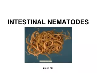
NEMATODES • Intestinal nematodes all mature into adults within the human intestinal tract. • Some species require an intermediate host to complete development
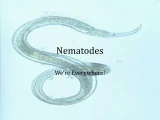
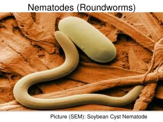
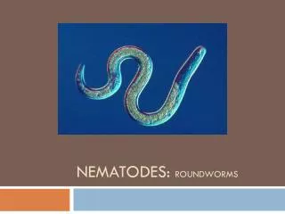
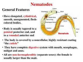
NEMATODES • The nematodes (nema: thread) are threadlike, nonsegmented parasites., • A few mm to 1m in length, with separated sexes. • They possess a complex tegument and a digestive tract.
NEMATODES • The males are usually smaller than the females and are equipped with copulatory organs that often show features specific to each species.
NEMATODES ●Development from the egg includes four larval stages and four moltings before the adult stage is reached. ●The larval forms of many of these roundworms may be distributed widely throughout the body
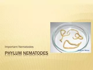
NEMATODES • Three of the intestinal nematodes are acquired by the ingestion of nematode eggs: ● Trichuris trichiura ("whipworm") ● Ascaris lumbricoides ● Enterobius vermicularis ("pinworm")
NEMATODES • Two worms are acquired when their larvae penetrate through the skin, usually of the foot: ● Necator americanus ("hookworm") ● Strongyloides stercoralis
NEMATODES • One is acquired by the ingestion of the encysted larvae in muscle (pork meat): Trichinella spiralis
NEMATODES Ascaris lumbricoides (Large Roundworm) Causative agent of “ascariosis”
Ascaris lumbricoides Occurrence ● The human large roundworm occurs worldwide. ● The main endemic regions, with prevalence rates of approx. 10–90%, include countries in Southeast Asia, Africa, and Latin America.
Ascaris lumbricoides Parasite and Lıfe Cycle ● The adult ascarids living in the small intestine are 15–40cm in length, about as thick as a pencil and of a yellowish pink color.
Ascaris lumbricoides ● The sexually mature females produce as many as 200 000 eggs per day, which are shed with feces in the unembryonated state.
The round-to-oval eggs are about 60 x 45 µm in size, have a thick, brownish shell and an uneven surface Ascaris lumbricoides
Ascaris lumbricoides At optimum temperatures of 20–25 ºC with sufficient moisture and oxygen, an infective larva in the egg develops within about three to six weeks
Ascaris lumbricoides Human infections result from peroral ingestion of eggs containing larvae, which hatch in the upper small intestine and penetrate into the veins of the intestinal wall.
Ascaris lumbricoides 1-They first migrate hematogenously “into the liver” 2- Then in four to seven days migrate “into the lungs “ where they leave the capillary network 3-Then migrate “into the alveoli”
Ascaris lumbricoides ● Via tracheopharyngeal migration they finally reach the digestive tract, where they further differentiate into adults in the small intestine. ● These periods lasts for seven to nine weeks. ● The lifespan of these parasites is 12–18 months.
Ascaris lumbricoides Epidemiology. Reservoir hosts of the parasite are humans. The excreted eggs remain viable for years in a moist environment (soil). *** Infective Ascaris eggs can be ingested by humans with contaminated foods, soil and, less frequently, in drinking water *** In endemic areas, the prevalence and intensity of A. lumbricoides infections are highest in children.
Ascaris lumbricoides Pathogenesis and clinical manifestations Mild infections frequently remain inapparent. In more severe infections, Larval migration in the lungs provoke hemorrhages and inflammatory infiltrations.
Ascaris lumbricoides During the intestinal phase of the infection, only some patients develop distinct clinical symptoms: abdominal discomfort with nausea, vomiting, pains,and diarrhea. Ascarids sometimes also migrate into the stomach, the pancreatic duct or the bile ducts and cause symptoms accordingly. Infection or frequent contact with volatile Ascaris antigens (laboratory staff!) can cause allergies.
Ascaris lumbricoides Diagnosis. ●An infection with sexually mature roundworms can be diagnosed by finding eggs in the stool ● Migrating Ascaris larvae can be indirectly detected by means of serological antibody detection (especially specific IgE), but this technique is seldom used in practice
Ascaris lumbricoides Therapy and control. Pyrantel, mebendazole, albendazole, and nitazoxanide are highly effective against the intestinal stages of Ascaris. Migratory stages are not affected by normal dosage levels. Due to the possibility of reinvasion of the intestine by larvae migrating in the body, the treatment should be repeated after two to three weeks.
Ascaris lumbricoides Preventive measures improvement of sanitation, good food hygiene practices (washing fruits and vegetables, cooking foods, etc.) and regular anthelminthic treatment of infected persons in endemic areas
Enterobius vermicularis(Pinworm) Causative agent of enterobiosis (oxyuriosis) Occurrence. The pinworm occurs in all parts of the world and is also a frequent parasite in temperate climate zones and developed countries. The age groups most frequently infected are five- to nine-year-old children and adults
Enterobius vermicularis Parasite and Lıfe Cycle Enterobius vermicularis which belongs to the Oxyurida has a white color. The males are 2–5mm long, The females 8–13 mm. The long, pointed tail of the female gives the pinworm its name.
Enterobius vermicularis Sexually mature pinworms live on the mucosa of the large intestine and lower small intestine. Following copulation, the males soon die off.
Enterobius vermicularis The females migrate to the anus, usually passing through the sphincter at night,then move about on the perianal skin, whereby each female lays about 10 000 eggs covered with a sticky proteinaceous layer enabling them to adhere to the skin.
Enterobius vermicularis • ● In severe infections, numerous living pinworms are often shed in stool and are easily recognizable as motile worms on the surface of the feces. • ● The eggs with their sticky surface they adhere to skin and other objects.
The eggs (about 50 - 30 µm in size) are slightly asymmetrical, ellipsoidal with thin shells Enterobius vermicularis
Enterobius vermicularis ● Freshly laid eggs contain an embryo that develops into an infective first-stage larva at skin temperature in about two days. ● Eggs that become detached from the skin remain viable for two to three weeks in a moist environment.
Enterobius vermicularis ● Infection occurs mainly by peroral uptake of eggs (each containing an infective larva) that are transmitted to the mouth with the fingers from the anal region or from various objects. ● The sticky eggs adhere to toys and items of everyday use or are disseminated with dust.
Enterobius vermicularis ● In the intestinal tract, larvae hatch from the ingested eggs, molt repeatedly, and develop into sexually mature pinworms in five to six weeks. ●“Retroinfection” is also conceivable, whereby infective larvae would be released at the anus to migrate back into the intestine.
Enterobius vermicularis Pathogenesis and clinical manifestations The pinworms living on the large intestine mucosa are fairly harmless. Occasionally, different stages of the pinwormb penetrate into the wall of the large intestine and the appendix or migrate into the vagina, uterus, fallopian tubes, and the abdominal cavity, where they cause inflammatory reactions.
Enterobius vermicularis ● The females of Enterobius produce in particular a very strong pruritus that may result in nervous disorders, developmental retardation, loss of weight and appetite, and other nonspecific symptoms. ● Scratch lesions and eczematous changes are produced in the anal area and can even spread to cover the entire skin.
Enterobius vermicularis Diagnosis. A tentative diagnosis based on clinical symptoms can be confirmed by detection of pinworms spontaneously excreted with feces and eggs adhering to the perianal skin ● Standard stool examination techniques are not sufficient to find the eggs. Egg detection by the “adhesive tape method” has proved most efficient
Enterobius vermicularis Therapy and prevention. albendazole, mebendazole and pyrantel are effective drugs. ● Reinfections are frequent, so that treatment usually should be repeated once or more times, extended to include all potential parasite carriers (e.g., family members, kindergarten members), and combined with measures, the purpose of which is to prevent egg dissemination.
Enterobius vermicularis ● Washing the perianal skin (especially in the morning), covering it with ointments, washing the hands, hot laundering of underwear, and cleaning contaminated objects with hot water
Enterobius vermicularis Thursday, April 30, 2009 Zeynep Simsek ,Ibrahim Koruk, Gülcan Gürses Harran University, Turkey Aysegul Cicek Copur Sanliurfa StateHospital, Turkey • Foodborne diseases continue to be one of the major public health problems in the world. 299 food-handlers working in Sanliurfa, Southeastern Anatolia (the response rate was 88.7%). Nasal and throat swab materials and stool samples were examined. The mean age of them was 26.7 (±9.6). Only 33.6% of the food-handlers had education above elementary school level. 52.2% of food handlers were suffering from intestinal parasites detected in the stools included Gardia lamblia (26.8%), Ascaris lumbricoides (10.7%), Tenya saginata (10%), Trichuris trichiura (1.3%), Enterobius Vermicularis(0.3%).
Enterobiosis in Sivas, Turkey from Past to Present, Effects on Primary School Children and Potential Risk Factors (4 located in City center, 4 in districts and 2 in villages) were classified as Region 1, Region 2 and Region 3, respectively and children completed a questionnaire about the potential risk factors. The overall egg positivity rate for E. vermicularis was 8.2% in Region 1, and the prevalence in the other regions was 7.0% and 14.8%, respectively. Children, aged 10–14 years, didn’t show a significantly higher egg positivity rate than younger children (0.10, p>0.05) and the infection rate for boys was not statistically different than girls Türkiye Parazitoloji Dergisi, 33 (1): 95 - 100, 2009 Serpil DEĞERLİ, Erdoğan MALATYALI, Semra ÖZÇELİK, Ali ÇELİKSÖZ Cumhuriyet Üniversitesi Tıp Fakültesi, Parazitoloji Bilim Dalı, Sivas, Türkiye
Trichuris trichiura (Whipworm)Causative agent of trichuriosis
Trichuris trichiura ● Trichuris trichiura occurs in humans and monkeys. ● Parasite has a worldwide distribution, it is found most frequently, like Ascaris lumbricoides, in moist, warm areas with low hygienic standards. ● The name whipworm characterizes the form of this 3–5cm long nematode with a very thin anterior part reminiscent of a whiplash and a thicker posterior “handle.”
Trichuristrichiura ●The adult nematodes live in the large intestine, mainly in the cecum. ● The females lay 2000–14 000 thick-shelled, yellow-brown eggs per day. ● The eggs are about 50–55 micromtr long and are readily identified by their lemonlike shape and hyaline polar plugs
Trichuristrichiura ●An infective larva develops in the egg within a few weeks. ● In moist surroundings, Trichuris eggs remain viable for months or even years.