Download
1 / 22
220 likes | 377 Vues
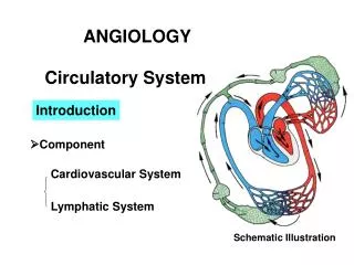
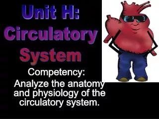
Unit H: Circulatory System. Competency: Analyze the anatomy and physiology of the circulatory system. Specific Objectives:. Explain the structure of the heart. STRUCTURES. Heart Arteries Veins Capillaries Blood and lymph are part of circulatory system.

E N D
Unit H: Circulatory System Competency: Analyze the anatomy and physiology of the circulatory system.
Specific Objectives: • Explain the structure of the heart.
STRUCTURES • Heart • Arteries • Veins • Capillaries • Blood and lymph are part of circulatory system http://faculty.plattsburgh.edu/david.curry/NUR464/Circul2.gif
SIZE SHAPE and LOCATION • Muscular organ • Size of a closed fist • Weighs 12-13 oz • Location – thoracic cavity • APEX – conical tip, lies on diaphragm, points left http://upload.wikimedia.org/wikipedia/commons/4/45/Illu_systemic_circuit.jpg
STETHOSCOPE an instrument used to hear (auscultate) the heartbeat. lubb dupp, lubb dupp, lubb dupp
PERICARDIUM pericardiumdouble layer of fibrous tissue that surrounds the heart http://www.nlm.nih.gov/medlineplus/ency/images/ency/fullsize/18081.jpg
Myocardium cardiac muscle tissue http://www.texasheart.org/HIC/Topics/images/myocard.jpg
Endocardium smooth inner lining of the heart The endocardium of this right ventricle is thickened, white, and glistening. http://www.texasheart.org/HIC/Topics/images/myocard.jpg
SEPTUM – partition (wall) that separates right half from left half http://www.columbiasurgery.org/pat/cardiac/img/novartis_212.gif
Superior and Inferior Vena Cava bring deoxygenated blood to the right atrium.
Pulmonary artery – takes blood away from the right ventricle to the lungs to pick up O2
Pulmonary veins – Bring Oxygenated blood from the lungs to the left Atrium. http://www.infomat.net/infomat/focus/health/health_curriculum/images/heart.gif
Aorta Takes the oxygenated blood away from the left ventricle to the rest of the body
Chambers and Valves SEPTUM – partition (wall) that separates right half from left half http://www.columbiasurgery.org/pat/cardiac/img/novartis_212.gif
Four heart valves permit flow of blood in one direction
TRICUSPID VALVE Between right atrium and right ventricle http://www.yourheart.org.uk/images/tricuspid_valve.jpg
BICUSPID (MITRAL) VALVEBetween left atrium and left ventricle http://www.yourheart.org.uk/images/mitral_valve.jpg
Semilunar valves are located where blood leaves the Heart. The PULMONARY SEMILUNAR VALVE & The AORTIC SEMILUNAR VALVE http://images.google.com/imgres?imgurl=http://www.skillstat.com/heartscape/heartValves.gif&imgrefurl=http://www.skillstat.com/heartscape/valves.htm&h=298&w=400&sz=20&hl=en&start=3&tbnid=M9Ji1QAnLlN9cM:&tbnh=92&tbnw=124&prev=/images%3Fq%3Dsemilunar%2Bvalves%26svnum%3D10%26hl%3Den%26lr%3D
Pretend you are a valve…have your thoughts flowing in one direction, toward studying for this huge test! • When is it ?