Download
1 / 65
650 likes | 982 Vues
Bone Physiology, Hormonal Control of Calcium Metabolism, and Osteoporosis. BONE PHYSIOLOGY. The skeleton has the following functions: The skeleton supports the body. The bones of the lower limbs support the entire body when we are standing, and the pelvic girdle supports the abdominal cavity.

E N D
Bone Physiology, Hormonal Control of Calcium Metabolism, and Osteoporosis
BONE PHYSIOLOGY The skeleton has the following functions: The skeleton supports the body. The bones of the lower limbs support the entire body when we are standing, and the pelvic girdle supports the abdominal cavity. The skeleton protects soft body parts. The bones of the skull protect the brain; the rib cage protects the heart and lungs. The skeleton produces blood cells. All bones in the fetus have red bone marrow that produces blood cells. In the adult, only certain bones produce blood cells. The skeleton stores minerals and fat. All bones have a matrix that contains calcium phosphate, a source of calcium ions and phosphate ions in the blood. Fat is stored in yellow bone marrow. The skeleton, along with the muscles, permits flexible body movement. While articulations (joints) occur between all the bones, we associate body movement in particular with the bones of the limbs. (Understanding Human Anatomy and Physiology, 5 Edition, p.84) Bone is a special form of connective tissue with a collagen framework impregnated with Ca2+ and PO43- salts, particularly hydroxyapatites. Old bone is constantly being resorbed and new bone formed, permitting remodeling that allows it to respond to the stresses and strains that are put upon it. It is a living tissue that is well vascularized and has a total blood flow of 200-400 mL/min in adult humans. (Review of Medical Physiology Ganong, 22 Edition)
Classification of bones; Long, short, flat, irregular, and round bones (Picture 1). (Understanding Human Anatomy and Physiology, 5 Edition, p.84) (Picture 2) A long bone, can be used to illustrate certain principles of bone anatomy. The bone is enclosed in a tough, fibrous, connective tissue covering called the periosteum, which is continuous with the ligaments and tendons that anchor bones. The periosteum contains blood vessels that enter the bone and service it cells. At both ends of a long bone is an expanded portion called an epiphysis; the portion between the epiphysis is called diaphysis. As shown in the adult bone in the picture, the diaphysis is not solid but has a medullary cavity containing yellow marrow. Yellow marrow contains large amounts of fat. The medullary cavity is bounded at the sides by cortical (compact) bone. The epiphyses contain trabecular (spongy) bone. Beyond the spongy bone is a thin shell of compact bone and, finally, a layer of hyaline cartilag called the articular cartilage. Articuar cartilage is so named because it occurs where bones articulate (join). Articulation is the joining together of bones at a joint. The medullary cavity and the space of spongy bone are line with endosteum, a thin, fibrous membrane. (Understanding Human Anatomy and Physiology, 5 Edition, p.84) In compact or cortical bone, which makes up the outer layer of most bones and accounts for 80% of the bone in the body; and trabecular or spongy bone inside the cortical bone, which makes up the remaining 20% of bone in the body. (Review of Medical Physiology Ganong, 22 Edition)
Compact bone contains many cylinder shaped units called osteons. The osteocytes (bone cells) are In tiny chambers called lacunae that occur between concentric layers of matrix called lamellae. The matrix contains collagenous protein fibers and mineral deposits, primarily of calcium and phosphorus salts. In each osteon, the lamellae and lacunae surround a single central canal. Blood vessels and nerves from the periosteum enter the central canal. The osteocytes have extensions that extend into passageways called canaliculi, and thereby the osteocytes are connected to each other and to the central canal. (Understanding Human Anatomy and Physiology, 5 Edition, p.84) Spongy bone contains numerous bony bars and plates, called trabeculae. Although lighter than compact bone, spongy bone is still designed for strength. Like braces used for support in buildings, the trabeculae of spongy bone follow lines of stress. (Understanding Human Anatomy and Physiology, 5 Edition, p.84) In compact bone, the surface-to-volume ratio is low, and bone cells lie in lacunae. They receive nutrients by way of canaliculi that ramify throughout the compact bone. Trabecullar bone is made up of spicules or plates, with a high surface-to-volume ratio and many cells sitting on the surface of the plates. Nutrients diffuse from bone ECF into the trabeculae, but in compact bone, nutrients are provided via haversian canals, which contain blood vessels. Around each haversian canal, collagen is arranged in concentric layers, forming cylinders called osteons or haversian systems (See also picture 3). (Review of Medical Physiology Ganong, 22 Edition)
The protein in bone matrix is over 90% type I collagen, which is also the major structural protein in tendons and skin. This collagen, which weight for weight is as strong as steel, is made up of a triple helix of three polypeptides bound tightly together. Two of these are identical α1 polypeptides encoded by one gene, and one is an α2 polypeptide encoded by a different gene. (Review of Medical Physiology Ganong, 22 Edition) The extracellular components of bone consist of a solid mineral phase in close association with an organic matrix, of which 90–95% is type I collagen . Single amino-acid substitutions in the helical portion of either the 1 (COL1A1) or 2 (COL1A2) chains of type I collagen disrupt the organization of bone in osteogenesisimperfecta. The severe skeletal fragility associated with these disorders highlights the importance of the fibrillar matrix in the structure of bone. (Harisson’s Principles of Internal Medicine, 17 Edition Volume 2, 2365-2366)
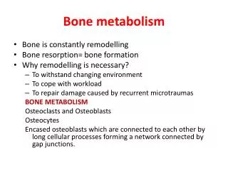
BONE REPAIR In the adult, bone is continually being broken down and built up again. Remodeling of bone is accomplished by two distinct cell types: osteoblasts produce bone matrix and osteoclastsresorb the matrix. The cells responsible for bone formation are osteoblasts and the cells responsible for bone resorption are osteoclasts.Osteoclasts derived from monocytes in red bone marrow break down bone, remove worn cells, and assist in depositing calcium in the blood. After a period of about three weeks, the osteoclasts disappear, and the bone is repaired by the work of osteoblasts. As they form new bone, osteoblasts take calcium from the blood. Eventually some of these cells get caught in the mineralized matrix they secrete and are converted to osteocytes, the cells found within the lacunae of osteons. See also picture 4; mechanism of bone remodelling and picture 5; schematic representation of bone remodelling. (Harisson’s Principles of Internal Medicine, 17 Edition Volume 2, 2365; Review of Medical Physiology Ganong, 22 Edition; and Understanding Human Anatomy and Physiology, 5 Edition, p.87) See picture 6; pathways regulating development of osteoblasts and osteoclast. (Harisson’s Principles of Internal Medicine, 17 Edition Volume 2, 2366) Osteoblasts are modified fibroblasts. Their early development from the mesenchyme is the same as that of fibroblasts, and the same large number of growth factors is involved. Later, ossification-specific factors begin to appear. One of the most interesting is the transcription factor Cbfa1. Mice in which the gene for Cbfa1 is knocked out develop to term with their skeletons made exclusively of cartilage; no ossification occurs. Normal osteoblasts are able to lay down type 1 collagen and form new bone. (Review of Medical Physiology Ganong, 22 Edition)
Osteoblasts synthesize and secrete the organic matrix. They are derived from cells of mesenchymal origin. Active osteoblasts are found on the surface of newly forming bone. As an osteoblast secretes matrix, which is then mineralized, the cell becomes an osteocyte, still connected with its blood supply through a series of canaliculi. Osteocytes comprise the vast majority of the cells in bone. Mineralization of the matrix, both in trabecular bone and in osteones of compact cortical bone (haversian systems), begins soon after the matrix is secreted (primary mineralization) but is not completed for several weeks or even longer (secondary mineralization). (Harisson’s Principles of Internal Medicine, 17 Edition Volume 2, 2366) Genetic studies in humans and mice have identified several key genes that control osteoblast development. Core-binding factor A1 (CBFA1, also called Runx2), is a transcription factor expressed specifically in chondrocyte (cartilage cells) and osteoblast progenitors, as well as in hypertrophic chondrocytes and mature osteoblasts. Runx2 regulates the expression of several important osteoblast proteins including osterix (another transcription factor needed for osteoblast maturation), osteopontin, bone sialoprotein, type I collagen, osteocalcin, and receptor-activator of NFB (RANK) ligand. Runx2 expression is regulated, in part, by bone morphogenic proteins (BMPs). Runx2-deficient mice are devoid of osteoblasts, whereas mice with a deletion of only one allele (Runx2 +/–) exhibit a delay in formation of the clavicles and some cranial bones. The latter abnormalities are similar to those in the human disorder cleidocranial dysplasia, which is also caused by heterozygous inactivating mutations in Runx2. (Harisson’s Principles of Internal Medicine, 17 Edition Volume 2, 2366)
The paracrine signaling molecule, Indian hedgehog (Ihh), also plays a critical role in osteoblast development, as evidenced by Ihh-deficient mice that lack osteoblasts in bone formed on a cartilage mold (endochondral ossification). Signals originating from members of the wnt (wingless-type mouse mammary tumor virus integration site) family of paracrine factors are also important. Humans and mice missing a wnt-family co-receptor, LRP5 (lipoprotein receptor–related protein 5), have osteoporosis. Remarkably, humans with an overactive form of LPR5 have increased bone mass. Numerous other growth-regulatory factors affect osteoblast function, including the three closely related transforming growth factor s, fibroblast growth factors (FGFs) 2 and 18, platelet-derived growth factor, and insulin-like growth factors (IGFs) I and II. Hormones such as parathyroid hormone (PTH) and 1,25-dihydroxyvitamin D [1,25(OH)2D] activate receptors expressed by osteoblasts to assure mineral homeostasis and to influence a variety of bone cell functions. (Harisson’s Principles of Internal Medicine, 17 Edition Volume 2, 2366) Osteoclasts, on the other hand, are members of the monocyte family. Stromal cells in the bone marrow, osteoblasts, and T lymphocytes all express a molecule called RANKL (RANK ligand) on their surface, and when they come in contact with appropriate monocytes they bind to RANKL receptors (RANK) on the surfaces of the monocytes.The combination converts the monocytes into osteoclasts. The precursor cells also secrete osteoprotegrin (OPG), which checks the conversion of the monocytes by competing with RANK for binding of RANKL (Picture 7). (Review of Medical Physiology Ganong, 22 Edition)
Resorption of bone is carried out mainly by osteoclasts, multinucleated cells that are formed by fusion of cells derived from the common precursor of macrophages and osteoclasts. Macrophage colony-stimulating factor (M-CSF) plays a critical role during several steps in the pathway and ultimately leads to fusion of osteoclast progenitor cells to form multinucleated, active osteoclasts. RANK ligand, a member of the tumor necrosis factor (TNF) family, is expressed on the surface of osteoblast progenitors and stromal fibroblasts. In a process involving cell-cell interactions, RANK ligand binds to the RANK receptor on osteoclast progenitors, stimulating osteoclast differentiation and activation. Alternatively, a soluble decoy receptor, referred to as osteoprotegerin, can bind RANK ligand and inhibit osteoclast differentiation. Several growth factors and cytokines (including interleukins 1, 6, and 11; TNF; and interferon ) modulate osteoclast differentiation and function. Most hormones that influence osteoclast function do not directly target this cell but instead influence M-CSF and RANK ligand signaling by osteoblasts. Both PTH and 1,25(OH)2D increase osteoclast number and activity, whereas estrogen decreases osteoclast number and activity by this indirect mechanism. Calcitonin, in contrast, binds to its receptor on the basal surface of osteoclasts and directly inhibits osteoclast function. (Harisson’s Principles of Internal Medicine, 17 Edition Volume 2, 2367)
Osteoclasts erode and absorb previously formed bone. They become attached to bone via integrins in a membrane extension called the sealing zone. This creates an isolated area between the bone and a portion of the osteoclast. Proton pumps, which are H+-dependent ATPases, then move from endosomes into the cell membrane apposed to the isolated area, and they acidify the area to approximately pH 4.0. The acidic pH dissolves hydroxyapatite, and acid proteases secreted by the cell break down collagen, forming a shallow depression in the bone (Picture 8). The products of digestion are then endocytosed and move across the osteoclast by transcytosis, with release into the interstitial fluid. The collagen breakdown products have pyridinoline structures, and pyridinolines can be measured in the urine as an index of the rate of bone resorption. (Review of Medical Physiology Ganong, 22 Edition) Osteoclast-mediated resorption of bone takes place in scalloped spaces (Howship's lacunae) where the osteoclasts are attached through a specific αvβ3integrin to components of the bone matrix such as osteopontin. The osteoclast forms a tight seal to the underlying matrix and secretes protons, chloride, and proteinases into a confined space likened to an extracellular lysosome. The active osteoclast surface forms a ruffled border that contains a specialized proton-pump ATPase, which secretes acid and solubilizes the mineral phase. Carbonic anhydrase (type II isoenzyme) within the osteoclast generates the needed protons. The bone matrix is resorbed in the acid environment adjacent to the ruffled border by proteases that act at low pH, such as cathepsin K. (Harisson’s Principles of Internal Medicine, 17 Edition Volume 2, 2367)
Throughout life, bone is being constantly resorbed and new bone is being formed. The calcium in bone turns over at a rate of 100% per year in infants and 18% per year in adults. Bone remodeling is mainly a local process carried out in small areas by populations of cells called bone-remodeling units. First, osteoclasts resorb bone, and then osteoblasts lay down new bone in the same general area. This cycle takes about 100 days. Modeling drifts also occur in which the shapes of bones change as bone is resorbed in one location and added in another. Osteoclasts tunnel into cortical bone followed by osteoblasts, whereas trabecular bone remodeling occurs on the surface of the trabeculae. About 5% of the bone mass is being remodeled by about 2 million bone-remodeling units in the human skeleton at any one time. The renewal rate for bone is about 4% per year for compact bone and 20% per year for trabecular bone. The remodeling is related in part to the stresses and strains imposed on the skeleton by gravity. (Review of Medical Physiology Ganong, 22 Edition) In adults, bone remodeling, and not modeling, is the principal metabolic skeletal process. Bone remodeling has two primary functions: (1) to repair microdamage within the skeleton to maintain skeletal strength, and (2) to supply calcium from the skeleton to maintain serum calcium. Remodeling may be activated by microdamage to bone as a result of excessive or accumulated stress. (Harisson’s Principles of Internal Medicine, 17 Edition Volume 2, 2399)
In addition, 4 steps of bone repair during fracture: • Hematoma. Within six to eight hours after a fracture, blood escapes from ruptured blood vessels and forms a hematoma (mass of clotted blood) in the space between the broken bones. • Fibrocartilaginous callus. Tissue repair begins, and fibrocartilage fills the space between the ends of the broken bone for about three weeks. • Bony callus. Osteoblasts produce trabeculae of spongy bone and convert the fibrocartilaginous callus to a bony callus that joins the broken bones together and lasts about three to four months. • Remodeling. Osteoblasts build new compact bone at the periphery, and osteoclasts reabsorb the spongy bone, creating a new medullary cavity. • Understanding Human Anatomy and Physiology, 5 Edition, p.87)
CALCIUM METABOLISM Three hormones are primarily concerned with the regulation of calcium metabolism. 1,25-Dihydroxycholecalciferol is a steroid hormone formed from vitamin D by successive hydroxylations in the liver and kidneys. Its primary action is to increase calcium absorption from the intestine. Parathyroid hormone (PTH) is secreted by the parathyroid glands. Its main action is to mobilize calcium from bone and increase urinary phosphate excretion. Calcitonin, a calcium-lowering hormone that in mammals is secreted primarily by cells in the thyroid gland, inhibits bone resorption. Although the role of calcitonin seems to be relatively minor, all three hormones probably operate in concert to maintain the constancy of the Ca2+ level in the body fluids. A fourth local hormone, parathyroid hormone-related protein (PTHrP), acts on one of the PTH receptors and is important in skeletal development in utero. Glucocorticoids, growth hormone, estrogens, and various growth factors also affect calcium metabolism. (Review of Medical Physiology Ganong, 22 Edition) The body of a young adult human contains about 1100 g (27.5 mol) of calcium. Ninety-nine percent of the calcium is in the skeleton. The plasma calcium, normally about 10 mg/dL (5 meq/L, 2.5 mmol/L), is partly bound to protein and partly diffusible. See distribution of calcium in normal human plasma. (Review of Medical Physiology Ganong, 22 Edition)
Over 99% of the 1–2 kg of calcium present normally in the adult human body resides in the skeleton, where it provides mechanical stability and serves as a reservoir sometimes needed to maintain extracellular fluid (ECF) calcium concentration. Skeletal calcium accretion first becomes significant during the third trimester of fetal life, accelerates throughout childhood and adolescence, reaches a peak in early adulthood, and gradually declines thereafter at rates that rarely exceed 1–2% per year. These slow changes in total skeletal calcium content contrast with relatively high daily rates of closely matched fluxes of calcium into and out of bone (~250–500 mg each), a process mediated by coupled osteoblastic and osteoclastic activity. Another 0.5–1% of skeletal calcium is freely exchangeable (e.g., in chemical equilibrium) with that in the ECF. (Harisson’s Principles of Internal Medicine, 17 Edition Volume 2, 2368) The calcium in bone is of two types: a readily exchangeable reservoir and a much larger pool of stable calcium that is only slowly exchangeable. Two independent but interacting homeostatic systems affect the calcium in bone. One is the system that regulates plasma Ca2+, and in the operation of this system, about 500 mmol of Ca2+ per day moves into and out of the readily exchangeable pool in the bone. The other system is the one concerned with bone remodeling by the constant interplay of bone resorption and deposition. However, the Ca2+ interchange between plasma and this stable pool of bone calcium is only about 7.5 mmol/d. (Review of Medical Physiology Ganong, 22 Edition) For further explanation of calcium homeostasis see picture 9 and picture 10.
The concentration of ionized calcium in the ECF must be maintained within a narrow range because of the critical role it plays in a wide array of cellular functions, especially those involved in neuromuscular activity, secretion, and signal transduction. Intracellular cytosolic free calcium levels are ~100 nmol/L and are 10,000-fold lower than ionized calcium concentration in the blood and ECF (1.1–1.3 mmol/L). This steep chemical gradient promotes rapid calcium influx through various membrane calcium channels that can be activated by hormones, metabolites, or neurotransmitters, swiftly changing cellular function. In blood, total calcium concentration is normally 2.2–2.6 mM (8.5–10.5 mg/dL), of which ~50% is ionized. The remainder is bound ionically to negatively charged proteins (predominantly albumin and immunoglobulins) or loosely complexed with phosphate, citrate, sulfate, or other anions. (Harisson’s Principles of Internal Medicine, 17 Edition Volume 2, 2368) A large amount of Ca2+ is filtered in the kidneys, but 98–99% of the filtered Ca2+ is reabsorbed. About 60% of the reabsorption occurs in the proximal tubules and the remainder in the ascending limb of the loop of Henle and the distal tubule. Distal tubular reabsorption is regulated by parathyroid hormone. (Review of Medical Physiology Ganong, 22 Edition)
HORMONAL CONTROL OF CALCIUM METABOLISM Control of the ionized calcium concentration in the ECF ordinarily is accomplished by adjusting the rates of calcium movement across intestinal and renal epithelia. These adjustments are mediated mainly via changes in blood levels of the hormones PTH and 1,25(OH)2D. Blood ionized calcium directly suppresses PTH secretion by activating parathyroid calcium-sensing receptors (CaSRs). Also, ionized calcium indirectly affects PTH secretion via effects on 1,25(OH)2D production. This active vitamin D metabolite inhibits PTH production by an incompletely understood mechanism of negative feedback. (Harisson’s Principles of Internal Medicine, 17 Edition Volume 2, 2368) Ca2+ is actively transported out of the intestine by a system in the brush border of the epithelial cells that involves a calcium-dependent ATPase, and this process is regulated by 1,25-dihydroxycholecalciferol. Some absorption also occurs by passive diffusion. When Ca2+ intake is high, 1,25-dihydroxycholecalciferol levels fall because of the increased plasma Ca2+. Consequently, Ca2+ absorption undergoes adaptation; i.e., it is high when the calcium intake is low and decreased when the calcium intake is high. Calcium absorption is also decreased by substances that form insoluble salts with Ca2+ (eg, phosphates and oxalates) or by alkalis, which favor formation of insoluble calcium soaps. A high-protein diet increases absorption in adults. (Review of Medical Physiology Ganong, 22 Edition)
Normal dietary calcium intake in the United States varies widely, ranging from 10–37 mmol/d (400–1500 mg/d). Many individuals, in an effort to prevent osteoporosis, routinely supplement this further with oral calcium salts to a total intake of 37–50 mmol/d (1500–2000 mg/d). Intestinal absorption of ingested calcium involves both active (transcellular) and passive (paracellular) mechanisms. Passive calcium absorption is nonsaturable and approximates 5% of daily calcium intake, whereas the active mechanism, controlled principally by 1,25(OH)2D, normally ranges from 20–70%. Active calcium transport occurs mainly in the proximal small bowel (duodenum and proximal jejunum), although some active calcium absorption occurs in most segments of the small intestine. Optimal rates of calcium absorption require gastric acid. This is especially true for weakly dissociable calcium supplements such as calcium carbonate. In fact, large boluses of calcium carbonate are poorly absorbed because of their neutralizing effect upon gastric acid. In achlorhydric subjects or for those taking drugs that inhibit gastric acid secretion, supplements should be taken with meals to optimize their absorption. Use of calcium citrate may be preferable in these circumstances. Calcium absorption may also be blunted in disease states such as pancreatic or biliary insufficiency, in which ingested calcium remains bound to unabsorbed fatty acids or other food constituents. At high levels of calcium intake, synthesis of 1,25(OH)2D is reduced, which decreases the rate of active intestinal calcium absorption. The opposite occurs with dietary calcium restriction. Some calcium, ~2.5–5.0 mmol/d (100–200 mg/d), is excreted as an obligate component of intestinal secretions and is not regulated by calciotropic hormones. (Harisson’s Principles of Internal Medicine, 17 Edition Volume 2, 2368)
The active transport of Ca2+ and PO43– from the intestine is increased by a metabolite of vitamin D. The term "vitamin D" is used to refer to a group of closely related sterols produced by the action of ultraviolet light on certain provitamins (Picture 11). Vitamin D3, which is also called cholecalciferol, is produced in the skin of mammals from 7-dehydrocholesterol by the action of sunlight. The reaction involves the rapid formation of previtamin D3, which is then converted more slowly to vitamin D3. Vitamin D3 and its hydroxylated derivatives are transported in the plasma bound to a globulin vitamin D-binding protein (DBP). Vitamin D3 is also ingested in the diet. (Review of Medical Physiology Ganong, 22 Edition) Vitamin D3 is metabolized by enzymes that are members of the cytochrome P450 (CYP) superfamily. In the liver, vitamin D3 is converted to 25-hydroxycholecalciferol (calcidiol, 25-OHD3). The 25-hydroxycholecalciferol is converted in the cells of the proximal tubules of the kidneys to the more active metabolite 1,25-dihydroxycholecalciferol, which is also called calcitriol or 1,25-(OH)2D3. 1,25-Dihydroxycholecalciferol is also made in the placenta, in keratinocytes in the skin, and in macrophages.The normal plasma level of 25-hydroxycholecalciferol is about 30 ng/mL, and that of 1,25-dihydroxycholecalciferol is about 0.03 ng/mL (approximately 100 pmol/L). The less active metabolite 24,25-dihydroxycholecalciferol is also formed in the kidneys.1,25-Dihydroxycholecalciferol is a hormone because it is produced in the body and transported in the bloodstream to produce effects in target cells. (Review of Medical Physiology Ganong, 22 Edition)
The mRNAs that are produced in response to 1,25-dihydroxycholecalciferol dictate the formation of a family of calbindin-D proteins. Calbindin-Ds are found in human intestine, brain, and kidneys and in many different tissues in rats. In the intestinal epithelium and many other tissues, two calbindins are induced: calbindin-D9K, and binds 2 Ca2+; and calbindin-D28K,and normally binds four Ca2+ even though it has six Ca2+-binding sites. In the intestine, increases in calbindin-D9K and calbindin-D28K levels are correlated with increased Ca2+ transport, but the precise way they facilitate Ca2+ movement across the intestinal epithelium is still uncertain. There is also evidence that 1,25-dihydroxycholecalciferol increases the number of Ca2+–H+ ATPase molecules in the intestinal cells; these are needed to pump Ca2+ into the interstitium.In addition to increasing Ca2+ absorption from the intestine, 1,25-dihydroxycholecalciferol facilitates Ca2+ reabsorption in the kidneys, increases the synthetic activity of osteoblasts, and is necessary for normal calcification of matrix. (Review of Medical Physiology Ganong, 22 Edition)
The formation of 25-hydroxycholecalciferol does not appear to be stringently regulated. However, the formation of 1,25-dihydroxycholecalciferol in the kidneys, which is catalyzed by 1α -hydroxylase, is regulated in a feedback fashion by plasma Ca2+ and PO43+ (Picture 12). Its formation is facilitated by PTH, and when the plasma Ca2+ level is low, PTH secretion is increased. When the plasma Ca2+ level is high, little 1,25-dihydroxycholecalciferol is produced, and the kidneys produce the relatively inactive metabolite 24,25- dihydroxycholecalciferol instead. This effect of Ca2+ on production of 1,25-dihydroxycholecalciferol is the mechanism that brings about adaptation of Ca2+ absorption from the intestine. The production of 1,25-dihydroxycholecalciferol is also increased by low and inhibited by high plasma PO43– levels, by a direct inhibitory effect of PO43– on 1α-hydroxylase. Additional control of 1,25-dihydroxycholecalciferol formation is exerted by a direct negative feedback effect of the metabolite on 1α -hydroxylase, a positive feedback action on the formation of 24,25-dihydroxycholecalciferol, and a direct action on the parathyroid gland to inhibit the production of mRNA for PTH. (Review of Medical Physiology Ganong, 22 Edition)
The normal plasma level of intact PTH is 10–55 pg/mL. The half-life of PTH is approximately 10 minutes, and the secreted polypeptide is rapidly cleaved by the Kupffer cells in the liver into midregion and carboxyl terminal fragments that are probably biologically inactive. PTH and these fragments are then cleared by the kidneys. PTH acts directly on bone to increase bone resorption and mobilize Ca2+. In addition to increasing the plasma Ca2+ and depressing the plasma phosphate, PTH increases phosphate excretion in the urine. This phosphaturic action is due to a decrease in reabsorption of phosphate in the proximal tubules. PTH also increases reabsorption of Ca2+ in the distal tubules, although Ca2+ excretion is often increased in hyperparathyroidism because the increase in the amount filtered overwhelms the effect on reabsorption. PTH also increases the formation of 1,25-dihydroxycholecalciferol, and this increases Ca2+ absorption from the intestine.On a longer timescale, PTH stimulates both osteoblasts and osteoclasts. (Review of Medical Physiology Ganong, 22 Edition)
Circulating ionized calcium acts directly on the parathyroid glands in a negative feedback fashion to regulate the secretion of PTH (Picture 13). The key to this regulation is a cell membrane Ca2+ receptor. This serpentine receptor is coupled via a G protein to phosphoinositide turnover and is found in many tissues. In the parathyroid, its activation inhibits PTH secretion. In this way, when the plasma Ca2+ level is high, PTH secretion is inhibited and the Ca2+ is deposited in the bones. When it is low, secretion is increased and Ca2+ is mobilized from the bones.1,25-Dihydroxycholecalciferol acts directly on the parathyroid glands to decrease preproPTH mRNA. Increased plasma phosphate stimulates PTH secretion by lowering plasma Ca2+ and inhibiting the formation of 1,25-dihydroxycholecalciferol. Magnesium is required to maintain normal parathyroid secretory responses. Impaired PTH release along with diminished target organ responses to PTH account for the hypocalcemia that occasionally occurs in magnesium deficiency. (Review of Medical Physiology Ganong, 22 Edition)
Secretion of calcitonin is increased when the thyroid gland is perfused with solutions containing a high Ca2+ concentration. Measurement of circulating calcitonin by immunoassay indicates that it is not secreted until the plasma calcium level reaches approximately 9.5 mg/dL and that above this calcium level, plasma calcitonin is directly proportionate to plasma calcium. β-Adrenergic agonists, dopamine, and estrogens also stimulate calcitonin secretion. Gastrin, CCK, glucagon, and secretin have all been reported to stimulate calcitonin secretion, with gastrin being the most potent stimulus. The plasma calcitonin level is elevated in Zollinger–Ellison syndromeand in pernicious anemia, in which the plasma gastrin level is also elevated. However, the dose of gastrin needed to stimulate calcitonin secretion produces an increase in plasma gastrin concentration greater than that produced by food, so it is premature to conclude that calcium in the intestine initiates secretion of a calcium-lowering hormone before the calcium is absorbed.Human calcitonin has a half-life of less than 10 minutes. (Review of Medical Physiology Ganong, 22 Edition)
Serpentine receptors for calcitonin are found in bones and the kidneys. Calcitonin lowers the circulating calcium and phosphate levels. It exerts its calcium-lowering effect by inhibiting bone resorption. This action is direct, and calcitonin inhibits the activity of osteoclasts in vitro. It also increases Ca2+ excretion in the urine.The exact physiologic role of calcitonin is uncertain. The calcitonin content of the human thyroid is low, and after thyroidectomy, bone density and plasma Ca2+ level are normal as long as the parathyroid glands are intact. Moreover, patients with medullary carcinoma of the thyroid have a very high circulating calcitonin level but no symptoms directly attributable to the hormone, and their bones are essentially normal. No syndrome due to calcitonin deficiency has been described. More hormone is secreted in young individuals, and it may play a role in skeletal development. It may protect against postprandial hypercalcemia. In addition, it may protect the bones of the mother from excess calcium loss during pregnancy. Bone formation in the infant and lactation are major drains on Ca2+ stores, and plasma concentrations of 1,25-dihydroxycholecalciferol are elevated in pregnancy. They would cause bone loss in the mother if bone resorption were not simultaneously inhibited by an increase in the plasma calcitonin level. (Review of Medical Physiology Ganong, 22 Edition)
The actions of the three principal hormones that regulate the plasma concentration of Ca2+ can now be summarized. PTH increases plasma Ca2+ by mobilizing this ion from bone. It increases Ca2+ reabsorption in the kidney, but this may be offset by the increase in filtered Ca2+. It also increases the formation of 1,25-dihydroxycholecalciferol. 1,25-Dihydroxycholecalciferol increases Ca2+ absorption from the intestine and increases Ca2+reabsorption in the kidneys. Calcitonin inhibits bone resorption and increases the amount of Ca2+ in the urine.Calcium metabolism is affected by various hormones in addition to 1,25-dihydroxycholecalciferol, PTH, PTHrP, and calcitonin. Glucocorticoids lower plasma Ca2+ levels by inhibiting osteoclast formation and activity, but over long periods they cause osteoporosis by decreasing bone formation and increasing bone resorption. They decrease bone formation by inhibiting protein synthesis in osteoblasts. They also decrease the absorption of Ca2+ and PO43– from the intestine and increase the renal excretion of these ions. This is why they depress the hypercalcemia of vitamin D intoxication. The decrease in plasma Ca2+ concentration increases the secretion of PTH, and bone resorption is facilitated. Growth hormone increases calcium excretion in the urine, but it also increases intestinal absorption of Ca2+, and this effect may be greater than the effect on excretion, with a resultant positive calcium balance. IGF-I generated by the action of growth hormone stimulates protein synthesis in bone. As noted above, thyroid hormones may cause hypercalcemia, hypercalciuria, and, in some instances, osteoporosis. Estrogens prevent osteoporosis by inhibiting the stimulatory effects of certain cytokines on osteoclasts. Insulin increases bone formation, and there is significant bone loss in untreated diabetes. (Review of Medical Physiology Ganong, 22 Edition)
Two subtypes of estrogens receptors (ERs), α and β, have been identified in bone and other tissues. Cells of monocyte lineage express both ER α and β, as do osteoblasts. Estrogen-mediated effects vary depending on the receptor type. Using ER knockout mouse models, elimination of ER α produces a modest reduction in bone mass, whereas mutation of ER β has less effect on bone. A male patient with a homozygous mutation of ER α had markedly decreased bone density as well as abnormalities in epiphyseal closure, confirming the important role of ER α in bone biology. The mechanism of estrogen action in bone through ERs is an area of active investigation (Picture 7). Although data are conflicting, estrogens may inhibit osteoclasts directly. However, the majority of estrogen (and androgen) effects on bone resorption are mediated indirectly through paracrine factors produced by osteoblasts. These actions include: (1) increasing IGF-I and TGF-, and (2) suppressing IL-1 ( and ), IL-6, TNF-, and osteocalcin synthesis. The indirect estrogen actions primarily decrease bone resorption. Estrogens regulate skeletal homeostasis in both men and women. Osteoporosis is due to increased bone resorption in both females and males and is associated with estrogen deficiency. (Harisson’s Principles of Internal Medicine, 17 Edition Volume 2, 2404-2405) (Deroo BJ et al: Estrogen receptors and human disease. J Clin Invest 116:561, 2006)
OSTEOPOROSIS: INTRODUCTION Osteoporosis, a condition characterized by decreased bone strength, is prevalent among postmenopausal women but also occurs in men and women with underlying conditions or major risk factors associated with bone demineralization. Its chief clinical manifestations are vertebral and hip fractures, although fractures can occur at any skeletal site. Osteoporosis affects >10 million individuals in the United States, but only a small proportion are diagnosed and treated. Osteoporosis is defined as a reduction in the strength of bone leading to an increased risk of fractures. Loss of bone tissue is associated with deterioration in skeletal microarchitecture. The World Health Organization (WHO) operationally defines osteoporosis as a bone density that falls 2.5 standard deviations (SD) below the mean for young healthy adults of the same gender—also referred to as a T-score of –2.5. Postmenopausal women who fall at the lower end of the young normal range (a T-score of >1 SD below the mean) are defined as having low bone density and are also at increased risk of osteoporosis. More than 50% of the fractures, including hip fractures, among postmenopausal women occur in this group. (Harisson’s Principles of Internal Medicine, 17 Edition Volume 2, 2397) The bone density criteria of World Health Organization are; normal (T-score of -1.0 or higher), osteopenia (T-score between -1.0 and -2.5), osteoporosis (T-score lower than -2.5). T-score is a comparison of a patient's bone density to that of a healthy thirty-year-old of the same sex and ethnicity. (WHO Scientific Group on the Prevention and Management of Osteoporosis (2000 : Geneva, Switzerland))
Cranney A, Jamal SA, Tsang JF, Josse RG, Leslie WD (2007). "Low bone mineral density and fracture burden in postmenopausal women. Canadian Medical Association Journal 177 (6): 575–80 In the United States, as many as 8 million women and 2 million men have osteoporosis (T-score < –2.5), and an additional 18 million individuals have bone mass levels that put them at increased risk of developing osteoporosis (e.g., bone mass T-score < –1.0). Osteoporosis occurs more frequently with increasing age as bone tissue is progressively lost. In women, the loss of ovarian function at menopause (typically about age 50) precipitates rapid bone loss such that most women meet the diagnostic criterion for osteoporosis by age 70–80. The epidemiology of fractures follows the trend for bone density loss. Fractures of the distal radius increase in frequency before age 50 and plateau by age 60, with only a modest age-related increase thereafter. In contrast, incidence rates for hip fractures double every 5 years after age 70 (Picture 14). This distinct epidemiology may be related to the way people fall as they age, with fewer falls on an outstretched hand and more falls directly on the hip. (Harisson’s Principles of Internal Medicine, 17 Edition Volume 2, 2397)
At least 1.5 million fractures occur each year in the United States as a consequence of osteoporosis. As the population continues to age, the total number of fractures will continue to escalate. (Harisson’s Principles of Internal Medicine, 17 Edition Volume 2, 2397) Lokasikejadianpatahtulang osteoporosis yang paling seringterjadiadalahpada vertebra, tulangleher femur, tulanggelangtangan (Colles). Adapunfrekuensipatahtulangleher femur adlaah 20% dari total jumlahpatahtulang osteoporosis. Di antarasemuapatahtulang osteoporosis, yang paling memberikanmasalahdibidangmorbiditas, mortalitas, bebansosioekonomik, dankualitashidupadalahpatahtulangleher femur. Bilatidakdiambiltindakanuntukmengatasi osteoporosis diperkirakanpadatahun 2050 jumlahpatahtulangleher femur diseluruhduniaakanmencapai 6,36 jutadanlebihdariseparuhnyadi Asia. Frekuensitertinggi osteoporosis wanita postmenopausal adalahpadausia 50-70 tahun. (Buku Ajar GangguanMuskuloskeletal, p.272) About 300,000 hip fractures occur each year in the United States, most of which require hospital admission and surgical intervention. The probability that a 50-year-old white individual will have a hip fracture during his or her lifetime is 14% for women and 5% for men; the risk for African Americans is lower (about half these rates). Hip fractures are associated with a high incidence of deep vein thrombosis and pulmonary embolism (20–50%) and a mortality rate between 5 and 20% during the year after surgery. (Harisson’s Principles of Internal Medicine, 17 Edition Volume 2, 2397)
There are about 700,000 vertebral crush fractures per year in the United States. Only a fraction of these are recognized clinically, since many are relatively asymptomatic and are identified incidentally during radiography for other purposes. Vertebral fractures rarely require hospitalization but are associated with long-term morbidity and a slight increase in mortality, primarily related to pulmonary disease. Multiple vertebral fractures lead to height loss (often of several inches), kyphosis, and secondary pain and discomfort related to altered biomechanics of the back. Thoracic fractures can be associated with restrictive lung disease, whereas lumbar fractures are associated with abdominal symptoms including distention, early satiety, and constipation. Approximately 250,000 wrist fractures occur in the United States each year. Fractures of other bones (estimated to be ~300,000 per year) also occur with osteoporosis, which is not surprising given that bone loss is a systemic phenomenon. Fractures of the pelvis and proximal humerus are clearly associated with osteoporosis. (Harisson’s Principles of Internal Medicine, 17 Edition Volume 2, 2397) In the United States and Europe, osteoporosis-related fractures are more common among women than men, presumably due to a lower peak bone mass as well as postmenopausal bone loss in women. Among individuals over the age of 50, any fracture should be considered as potentially related to osteoporosis, irrespective of the circumstances of fracture. Osteoporotic bone is more likely to fracture than normal bone at any level of trauma, and a fracture in a person over 50 should trigger evaluation for osteoporosis. (Harisson’s Principles of Internal Medicine, 17 Edition Volume 2, 2398)
OSTEOPOROSIS: RISK FACTORS Although some fractures are the result of major trauma, the threshold for fracture is reduced for an osteoporotic bone (Picture 15). In addition to bone density there are a number of risk factors for fracture; the common ones are summarized. Chronic diseases with inflammatory components that increase skeletal remodeling, such as rheumatoid arthritis, increase the risk of osteoporosis, as do diseases associated with malabsorption. Chronic diseases that increase the risk of falling or frailty, including dementia, Parkinson's disease, and multiple sclerosis, also increase fracture risk. (Harisson’s Principles of Internal Medicine, 17 Edition Volume 2, 2397-2398) Beberapafaktorpredisposisi osteoporosis, sebagaiberikut: Gangguanendokrin (hiperparatiroidism, hipogonadism, diabetes melitus, cushing syndrome, prolaktinoma, akromegali Gangguannutrisidan gastrointestinal (inflammatory bowel disease, celiac disease, malnutrisi, gastric bypass, chronic liver disease Penyakitginjal (gagalginjalkronik, hiperkalsiuria) Rematik (rheumatoid arthritis, lupus, ankylosingspondylitis) Gangguanhematologi (thalasemia, multiple myeloma, leukimia, lifoma, hemofilia) Genetik (Ehlers-Danlos syndrome, Marfan syndrome, osteogenesisimperfekta) Drugs (Kortikosteroid [>5mg/hari minimal pemberian 3 bulan], Antikonvulsan, kemoterapik/obat-obatantransplantasi [cyclosporine, methotrexate], lithium, aromatase inhibitor [exemestane, anastrozole]) (Buku Ajar GangguanMuskuloskeletal, p.272)
Various genetic and acquired diseases are associated with an increase in the risk of osteoporosis. Mechanisms that contribute to bone loss are unique for each disease and typically result from multiple factors including nutrition, reduced physical activity levels, and factors that affect bone-remodeling rates. (Harisson’s Principles of Internal Medicine, 17 Edition Volume 2, 2400) Osteoporosis has multiple causes, but by far the commonest form is involutional osteoporosis. All normal humans gain bone early in life, during growth. After a plateau, they begin to lose bone as they grow older (Picture 16). When this loss is accelerated or exaggerated, it leads to osteoporosis.Adult women have less bone mass than adult men, and after menopause they initially lose it more rapidly than men of comparable age do. Consequently, they are more prone to development of serious osteoporosis. The cause of the bone loss after menopause is primarily estrogen deficiency, and estrogen treatment arrests the progress of the disease. Estrogens inhibit secretion of cytokines such as IL-1, IL-6, and TNFα , and these cytokines foster the development of osteoclasts. Estrogen also stimulates production of TGF-β , and this cytokine increases apoptosis of osteoclasts. However, it now appears that even small doses of estrogens may increase the incidence of uterine and breast cancer, and in carefully controlled studies, estrogens do not protect against cardiovascular disease. Therefore, the decision to treat a postmenopausal woman with estrogens depends on a careful weighing of the risk–benefit ratio. (Review of Medical Physiology Ganong, 22 Edition)
Peak bone mass may be impaired by inadequate calcium intake during growth among other nutritional factors (calories, protein, and other minerals), thereby leading to increased risk of osteoporosis later in life. During the adult phase of life, insufficient calcium intake contributes to relative secondary hyperparathyroidism and an increase in the rate of bone remodeling to maintain normal serum calcium levels. PTH stimulates the hydroxylation of vitamin D in the kidney, leading to increased levels of 1,25-dihydroxyvitamin D [1,25(OH)2D] and enhanced gastrointestinal calcium absorption. PTH also reduces renal calcium loss. Although these are all appropriate compensatory homeostatic responses for adjusting calcium economy, the long-term effects are detrimental to the skeleton because the increased remodeling rates and the ongoing imbalance between resorption and formation at remodeling sites combine to accelerate loss of bone tissue. (Harisson’s Principles of Internal Medicine, 17 Edition Volume 2, 2399) In women living in northern latitudes, it has been shown that vitamin D levels decline during the winter months. This is associated with seasonal bone loss, reflecting increased bone turnover. Treatment with vitamin D can return levels to normal [>75 mol/L (30 ng/mL)] and prevent the associated increase in bone remodeling, bone loss, and fractures. Reduced fracture rates have also been documented among individuals in northern latitudes who have greater vitamin D intake and have higher 25(OH)D levels. (Harisson’s Principles of Internal Medicine, 17 Edition Volume 2, 2400)
Inactivity, such as prolonged bed rest or paralysis, results in significant bone loss. Concordantly, athletes have higher bone mass than the general population. These changes in skeletal mass are most marked when the stimulus begins during growth and before the age of puberty. Adults are less capable than children of increasing bone mass following restoration of physical activity. Fracture risk is lower in rural communities and in countries where physical activity is maintained into old age. However, when exercise is initiated during adult life, the effects of moderate exercise on the skeleton are modest, with a bone mass increase of 1–2% in short-term studies of <2 years' duration. It is argued that more active individuals are less likely to fall and are more capable of protecting themselves upon falling, thereby reducing fracture risk. (Harisson’s Principles of Internal Medicine, 17 Edition Volume 2, 2400) A large number of medications used in clinical practice have potentially detrimental effects on the skeleton (glucocorticoid, Cyclosporine, Anticonvulsants, Aromatase inhibitors, Heparin, Gonadotropin releasing hormone agonists, Lithium). Glucocorticoids are the most common cause of medication-induced osteoporosis. It is often not possible to determine the extent to which osteoporosis is related to the glucocorticoid or to other factors, as treatment is superimposed on the effects of the primary disease, which may in itself be associated with bone loss (e.g., rheumatoid arthritis). Aromatase inhibitors, used in various stages for breast cancer treatment, have also been shown to have a detrimental effect on bone density and risk of fracture. (Harisson’s Principles of Internal Medicine, 17 Edition Volume 2, 2400-2401)
Osteoporotic fractures are a well-characterized consequence of the hypercortisolism associated with Cushing's syndrome. However, the therapeutic use of glucocorticoids is by far the most common form of glucocorticoid-induced osteoporosis. Glucocorticoids are widely used in the treatment of a variety of disorders, including chronic lung disorders, rheumatoid arthritis and other connective tissue diseases, inflammatory bowel disease, and posttransplantation. Osteoporosis and related fractures are serious side effects of chronic glucocorticoid therapy. Because the effects of glucocorticoids on the skeleton are often superimposed upon the consequences of aging and menopause, it is not surprising that women and the elderly are most frequently affected. The skeletal response to steroids is remarkably heterogeneous, however, and even young, growing individuals treated with glucocorticoids can present with fractures. (Harisson’s Principles of Internal Medicine, 17 Edition Volume 2, 2407) The risk of fractures depends on the dose and duration of glucocorticoid therapy, although recent data suggest that there may be no completely safe dose. Bone loss is more rapid during the early months of treatment, and trabecular bone is more severely affected than cortical bone. As a result, fractures have been shown to increase within 3 months of steroid treatment. There is an increase in fracture risk in both the axial and appendicular skeleton, including risk of hip fracture. Bone loss can occur with any route of steroid administration including high-dose inhaled glucocorticoids and intraarticular injections (Harisson’sPrinciples of Internal Medicine, 17 Edition Volume 2, 2407)
Glucocorticoids increase bone loss by multiple mechanisms including: (1) inhibition of osteoblast function and an increase in osteoblast apoptosis, resulting in impaired synthesis of new bone; (2) stimulation of bone resorption, probably as a secondary effect; (3) impairment of the absorption of calcium across the intestine, probably by a vitamin D–independent effect; (4) increase of urinary calcium loss and perhaps induction of some degree of secondary hyperparathyroidism; (5) reduction of adrenal androgens and suppression of ovarian and testicular secretion of estrogens and androgens; and (6) induction of glucocorticoidmyopathy, which may exacerbate effects on skeletal and calcium homeostasis as well as increase the risk of falls. (Harisson’s Principles of Internal Medicine, 17 Edition Volume 2, 2407-2408) The use of cigarettes over a long period has detrimental effects on bone mass. These effects may be mediated directly, by toxic effects on osteoblasts, or indirectly by modifying estrogen metabolism. On average, cigarette smokers reach menopause 1–2 years earlier than the general population. Cigarette smoking also produces secondary effects that can modulate skeletal status, including intercurrent respiratory and other illnesses, frailty, decreased exercise, poor nutrition, and the need for additional medications (e.g., glucocorticoids for lung disease). (Harisson’s Principles of Internal Medicine, 17 Edition Volume 2, 2401)
OSTEOPOROSIS: PATHOPHYSIOLOGY Osteoporosis is caused by a relative excess of osteoclastic function. Loss of bone matrix in this condition (Picture 17) is marked, and the incidence of fractures is increased. Fractures are particularly common in the distal forearm (Colles' fracture), vertebral body, and hip. All of these areas have a high content of trabecular bone, and since trabecular bone is more active metabolically, it is lost more rapidly. Fractures of the vertebrae with compression cause kyphosis, with the production of a typical "widow's hump/Dowager hump“(Picture 18) that is common in elderly women with osteoporosis. Fractures of the hip in elderly individuals are associated with a mortality rate of 12–20%, and half of those who survive require prolonged expensive care. (Review of Medical Physiology Ganong, 22 Edition) In young adults resorbed bone is replaced by an equal amount of new bone tissue. Thus, the mass of the skeleton remains constant after peak bone mass is achieved in adulthood. After age 30–45, however, the resorption and formation processes become imbalanced, and resorption exceeds formation. This imbalance may begin at different ages and varies at different skeletal sites; it becomes exaggerated in women after menopause. Excessive bone loss can be due to an increase in osteoclastic activity and/or a decrease in osteoblastic activity. In addition, an increase in remodeling activation frequency, and thus the number of remodeling sites, can magnify the small imbalance seen at each remodeling unit. (Harisson’s Principles of Internal Medicine, 17 Edition Volume 2, 2399)
Increased recruitment of bone remodeling sites produces a reversible reduction in bone tissue but can also result in permanent loss of tissue and disrupted skeletal architecture. In trabecular bone, if the osteoclasts penetrate trabeculae, they leave no template for new bone formation to occur and, consequently, rapid bone loss ensues and cancellous connectivity becomes impaired. A higher number of remodeling sites increases the likelihood of this event. In cortical bone, increased activation of remodeling creates more porous bone. The effect of this increased porosity on cortical bone strength may be modest if the overall diameter of the bone is not changed. However, decreased apposition of new bone on the periosteal surface coupled with increased endocorticalresorption of bone decreases the biomechanical strength of long bones. Even a slight exaggeration in normal bone loss increases the risk of osteoporosis-related fractures, due to the architectural changes that occur. (Harisson’s Principles of Internal Medicine, 17 Edition Volume 2, 2399)
OSTEOPOROSIS: DIAGNOSIS Several noninvasive techniques are now available for estimating skeletal mass or density. These include dual-energy x-ray absorptiometry (DXA), single-energy x-ray absorptiometry (SXA), quantitative CT, and ultrasound. DXA is a highly accurate x-ray technique that has become the standard for measuring bone density in most centers. Though it can be used for measurements of any skeletal site, clinical determinations are usually made of the lumbar spine and hip. All of these techniques for measuring BMD have been approved by the U.S. Food and Drug Administration (FDA) based upon their capacity to predict fracture risk. In younger individuals, such as perimenopausal or early postmenopausal women, spine measurements may be the most sensitive indicator of bone loss. (Harisson’s Principles of Internal Medicine, 17 Edition Volume 2, 2401) Clinical guidelines have been developed for use of bone densitometry in clinical practice. The original National Osteoporosis Foundation guidelines recommend bone mass measurements in postmenopausal women, assuming they have one or more risk factors for osteoporosis in addition to age, gender, and estrogen deficiency. The guidelines further recommend that bone mass measurement be considered in all women by age 65, a position ratified by the U.S. Preventive Health Services Task Force. Criteria approved for Medicare reimbursement of BMD are summarized. (Harisson’s Principles of Internal Medicine, 17 Edition Volume 2, 2401)
OSTEOPOROSIS: ROUTINE LABORATORY EVALUATION There is no established algorithm for the evaluation of women presenting with osteoporosis. A general evaluation that includes complete blood count, serum and 24-h urine calcium, and renal and hepatic function tests is useful for identifying selected secondary causes of low bone mass, particularly for women with fractures. (Harisson’s Principles of Internal Medicine, 17 Edition Volume 2, 2402) An elevated serum calcium level suggests hyperparathyroidism or malignancy, whereas a reduced serum calcium level may reflect malnutrition and osteomalacia. In the presence of hypercalcemia, a serum PTH level differentiates between hyperparathyroidism and malignancy. A low urine calcium (<50 mg/24 h) suggests osteomalacia, malnutrition, or malabsorption; a high urine calcium (>300 mg/24 h) is indicative of hypercalciuria and must be investigated further. Hypercalciuria occurs primarily in three situations: (1) a renal calcium leak, which is more frequent in males with osteoporosis; (2) absorptive hypercalciuria, which can be idiopathic or associated with increased 1,25(OH)2D in granulomatous disease; or (3) hematologic malignancies or conditions associated with excessive bone turnover such as Paget's disease, hyperparathyroidism, and hyperthyroidism. (Harisson’s Principles of Internal Medicine, 17 Edition Volume 2, 2402)
Individuals who have osteoporosis-related fractures or bone density in the osteoporotic range should have a measurement of serum 25(OH)D level, since the intake of vitamin D required to achieve a target level >32 ng/mL is very variable. Vitamin D levels should be optimized in all individuals being treated for osteoporosis. Hyperthyroidism should be evaluated by measuring thyroid-stimulating hormone (TSH). (Harisson’s Principles of Internal Medicine, 17 Edition Volume 2, 2402) When there is clinical suspicion of Cushing's syndrome, urinary free cortisol levels or a fasting serum cortisol should be measured after overnight dexamethasone. When bowel disease, malabsorption, or malnutrition is suspected, serum albumin, cholesterol, and a complete blood count should be checked. Asymptomatic malabsorption might be heralded by anemia (macrocytic–vitamin B12 or folate deficiency; or microcytic-iron deficiency) or low serum cholesterol or urinary calcium levels. If these or other features suggest malabsorption, further evaluation is required. Asymptomatic celiac disease with selective malabsorption is being found with increasing frequency; the diagnosis can be made by testing for antigliadin, antiendomysial, or transglutaminase antibodies but may require endoscopic biopsy. When osteoporosis is found associated with symptoms of rash, multiple allergies, diarrhea, or flushing, mastocytosis should be excluded using 24-h urine histamine collection or serum tryptase. (Harisson’s Principles of Internal Medicine, 17 Edition Volume 2, 2402)
Because of the prevalence of glucocorticoid-induced bone loss, it is important to evaluate the status of the skeleton in all patients starting or already receiving long-term glucocorticoid therapy. Modifiable risk factors should be identified, including those for falls. Examination should include height and muscle strength testing. Laboratory evaluation should include an assessment of 24-h urinary calcium. All patients on long-term (>3 months) glucocorticoids should have measurement of bone mass at both the spine and hip using DXA. If only one skeletal site can be measured, it is best to assess the spine in individuals <60 years and the hip for those >60 years. (Harisson’s Principles of Internal Medicine, 17 Edition Volume 2, 2408) Myeloma can masquerade as generalized osteoporosis, although it more commonly presents with bone pain and characteristic "punched-out" lesions on radiography. Serum and urine electrophoresis and evaluation for light chains in urine are required to exclude this diagnosis. A bone marrow biopsy may be required to rule out myeloma (in patients with equivocal electrophoretic results) and can also be used to exclude mastocytosis, leukemia, and other marrow infiltrative disorders, such as Gaucher's disease. (Harisson’s Principles of Internal Medicine, 17 Edition Volume 2, 2402)
Several biochemical tests are now available that provide an index of the overall rate of bone remodeling. Biochemical markers are usually characterized as those related primarily to bone formation or bone resorption. For the most part, remodeling markers do not predict rates of bone loss well enough to use this information clinically. However, markers of bone resorption may help in the prediction of fracture risk, independently of bone density, particularly in older individuals. In women 65 years, when bone density results are greater than the usual treatment thresholds noted above, a high level of bone resorption should prompt consideration of treatment. The primary use of biochemical markers is for monitoring the response to treatment. With the introduction of antiresorptive therapeutic agents, bone remodeling declines rapidly, with the fall in resorption occurring earlier than the fall in formation. Inhibition of bone resorption is maximal within 3–6 months. A decline in resorptive markers can be ascertained after treatment with bisphosphonates or estrogen; this effect is less marked after treatment with either raloxifene or intranasal calcitonin. (Harisson’s Principles of Internal Medicine, 17 Edition Volume 2, 2402)
OSTEOPOROSIS: NON-PHARMACOLOGIC AND NUTRITIONAL TREATMENTS Most guidelines suggest that patients be considered for treatment when BMD is >2.5 SD below the mean value for young adults (T-score –2.5), a level consistent with the diagnosis of osteoporosis. Treatment should also be considered in postmenopausal women with risk factors, even if BMD is not in the osteoporosis range. It is important to consider the risk of fracture for individuals, including those whose BMD is within the premenopausal range. Risk factors (age, prior fracture, family history of hip fracture, low body weight, cigarette consumption, excessive alcohol, steroid use, and rheumatoid arthritis) can be combined with BMD to assess the likelihood of a fracture over a 5- or 10-year period. (Harisson’s Principles of Internal Medicine, 17 Edition Volume 2, 2401) Although osteoporosis indicates a high likelihood of fracture, many fragility fractures occur in people with bone density values above the defined level. Fractures can be better predicted by adding clinical risk factors that contribute to fracture risk independently of bone mineral density. This approach is being developed under the auspices of the WHO and will be delivered in the form of an algorithm that enables the probability of a fracture to be calculated from clinical risk factors with or without bone mineral density values. Intervention thresholds based on cost effectiveness can be used to make a decision about treatment. (Poole KE, Compston JE: Osteoporosis and its management. BMJ 333:1251, 2006)
Treatment of the patient with osteoporosis frequently involves management of acute fractures as well as treatment of the underlying disease. Hip fractures almost always require surgical repair if the patient is to become ambulatory again. Other fractures (e.g., vertebral, rib, and pelvic fractures) are usually managed with supportive care, requiring no specific orthopedic treatment. For acutely symptomatic fractures, treatment with analgesics is required, including NSAID’s and/or acetaminophen, sometimes with the addition of a narcotic agent (codeine or oxycodone). Short periods of bed rest may be helpful for pain management, but, in general, early mobilization is recommended as it helps prevent further bone loss associated with immobilization. Multiple vertebral fractures are often associated with psychological symptoms. The changes in body configuration and back pain can lead to marked loss of self-image and a secondary depression. Medication may be necessary when depressive features are present. (Harisson’s Principles of Internal Medicine, 17 Edition Volume 2, 2402-2403)
Patients should be thoroughly educated to reduce the impact of modifiable risk factors associated with bone loss and falling. Medications should be reviewed to ensure that all are necessary. Glucocorticoid medication, if present, should be evaluated to determine that it is truly indicated and is being given in doses as low as possible. For those on thyroid hormone replacement, TSH testing should be performed to determine that an excessive dose is not being used, as thyrotoxicosis can be associated with increased bone loss. If nocturia occurs, the frequency should be reduced, if possible (e.g., by decreasing or modifying diuretic use). Elderly patients with neurologic impairment (e.g., stroke, Parkinson's disease, Alzheimer's disease) are particularly at risk of falling and require specialized supervision and care. (Harisson’s Principles of Internal Medicine, 17 Edition Volume 2, 2403) A large body of data indicates that optimal calcium intake reduces bone loss and suppresses bone turnover. The preferred source of calcium is from dairy products and other foods, but many patients require calcium supplementation. Food sources of calcium are dairy products (milk, yogurt, and cheese) and fortified foods such as certain cereals, waffles, snacks, juices, and crackers. Some of these fortified foods contain as much calcium per serving as milk. If a calcium supplement is required, it should be taken in doses <600 mg at a time, as the calcium absorption fraction decreases at higher doses. Calcium supplements should be calculated based on the elemental calcium content of the supplement, not the weight of the calcium salt. Calcium supplements containing carbonate are best taken with food since they require acid for solubility. Calcium citrate supplements can be taken at any time. (Harisson’s Principles of Internal Medicine, 17 Edition Volume 2, 2403)
Although side effects from supplemental calcium are minimal (eructation and constipation mostly with carbonate salts), individuals with a history of kidney stones should have a 24-h urine calcium determination before starting increased calcium to avoid significant hypercalciuria. (Harisson’s Principles of Internal Medicine, 17 Edition Volume 2, 2403) Large segments of the population do not obtain sufficient vitamin D to maintain what is now considered an adequate supply [serum 25(OH)D consistently >75μmol/L (30 ng/mL)]. Since vitamin D supplementation at doses that would achieve these serum levels is safe and inexpensive, the Institute of Medicine recommends daily intakes of 200 IU for adults <50 years of age, 400 IU for those from 50–70 years, and 600 IU for those >70 years. Multivitamin tablets usually contain 400 IU, and many calcium supplements also contain vitamin D. Some data suggest that higher doses (>1000 IU) may be required in the elderly and chronically ill. (Harisson’s Principles of Internal Medicine, 17 Edition Volume 2, 2403) Several controlled clinical trials of calcium plus vitamin D have confirmed reductions in clinical fractures, including fractures of the hip (~20–30% risk reduction). (Harisson’s Principles of Internal Medicine, 17 Edition Volume 2, 2403)
Exercise in young individuals increases the likelihood that they will attain the maximal genetically determined peak bone mass. Meta-analyses of studies performed in postmenopausal women indicate that weight-bearing exercise prevents bone loss but does not appear to result in substantial gain of bone mass. This beneficial effect wanes if exercise is discontinued. Most of the studies are short-term, and a more substantial effect on bone mass is likely if exercise is continued over a long period of time. Exercise also has beneficial effects on neuromuscular function, and it improves coordination, balance, and strength, thereby reducing the risk of falling. A walking program is a practical way to start. Other activities such as dancing, racquet sports, cross-country skiing, and use of gym equipment are also recommended, depending on the patient's personal preference and general condition. Even women who cannot walk benefit from swimming or water exercises, not so much for the effects on bone, which are quite minimal, but because of effects on muscle. Exercise habits should be consistent, optimally at least three times a week. (Harisson’s Principles of Internal Medicine, 17 Edition Volume 2, 2404) Bone loss caused by glucocorticoids can be prevented, and the risk of fractures significantly reduced. Strategies must include using the lowest dose of glucocorticoid for disease management. Topical and inhaled routes of administration are preferred, where appropriate. Risk factor reduction is important, including smoking cessation, limitation of alcohol consumption, and participation in weight-bearing exercise, when appropriate. All patients should receive an adequate calcium and vitamin D intake from the diet or from supplements. (Harisson’s Principles of Internal Medicine, 17 Edition Volume 2, 2408)
OSTEOPOROSIS: PHARMACOLOGIC TREATMENTS Therapeutic options for osteoporosis have increased considerably over recent years. Until fairly recently, estrogen treatment, either by itself or in concert with a progestin, was the primary therapeutic agent for prevention or treatment of osteoporosis. A large body of clinical trial data indicates that various types of estrogens (conjugated equine estrogens, estradiol, estrone, esterified estrogens, ethinylestradiol, and mestranol) reduce bone turnover, prevent bone loss, and induce small increases in bone mass of the spine, hip, and total body. The effects of estrogen are seen in women with natural or surgical menopause and in late postmenopausal women with or without established osteoporosis. Estrogens are efficacious when administered orally or transdermally. One large study, referred to as PEPI (Postmenopausal Estrogen/Progestin Intervention Trial), indicated that C-21 progestins alone do not augment the effect of standard estrogen doses on bone mass (Picture 19). (Harisson’s Principles of Internal Medicine, 17 Edition Volume 2, 2404) For oral estrogens, the standard recommended doses have been 0.3 mg/d for esterified estrogens, 0.625 mg/d for conjugated equine estrogens, and 5μg/d for ethinylestradiol. For transdermal estrogen, the commonly used dose supplies 50μg estradiol per day, but a lower dose may be appropriate for some individuals. Dose response data for conjugated equine estrogens indicate that lower doses (0.3 and 0.45 mg/d) are effective. Doses even lower have been associated with bone mass protection. (Harisson’s Principles of Internal Medicine, 17 Edition Volume 2, 2404)
Epidemiologic databases indicate that women who take estrogen replacement have a 50% reduction, on average, of osteoporotic fractures, including hip fractures. The beneficial effect of estrogen is greatest among those who start replacement early and continue the treatment; the benefit declines after discontinuation such that there is no residual protective effect against fracture by 10 years after discontinuation. The estrogen-progestin arm of the WHI in >16,000 postmenopausal healthy women indicated that hormone therapy reduces the risk of hip and clinical spine fracture by 34% and all clinical fractures by 24%. (Harisson’s Principles of Internal Medicine, 17 Edition Volume 2, 2404) In many trials and studies, estrogens use shows higher risk to develop breast cancer, ovarian cancer, endometrial cancer (in the absence of sufficient progesterone [the “unopposed estrogen hypothesis”], and this hypothesis has been supported by many studies), and venous thromboembolic events. (Deroo BJ et al: Estrogen receptors and human disease. J Clin Invest 116:561, 2006) In contrast, estrogens use shows lower risk to develop colon cancer, osteoporosis, parkinsondisease, and alzheimer disease. clinical studies also support a protective role for estrogens in the regulation of lipid and cholesterol levels, Estrogen is known to increase HDL plasma levels and decreased LDL plasma levels; however, this response varies greatly in women and may be due in part to genetic factors. (Deroo BJ et al: Estrogen receptors and human disease. J Clin Invest 116:561, 2006)