
Exploring the Layers of Skin Anatomy: Epidermis and Dermis
Dive into the intricate structure of the skin, detailed layers, and functions including epidermis, dermis, and subcutaneous layer. Learn about skin cells, growth, skin color factors, and tissue repair process.

Exploring the Layers of Skin Anatomy: Epidermis and Dermis
E N D
Presentation Transcript
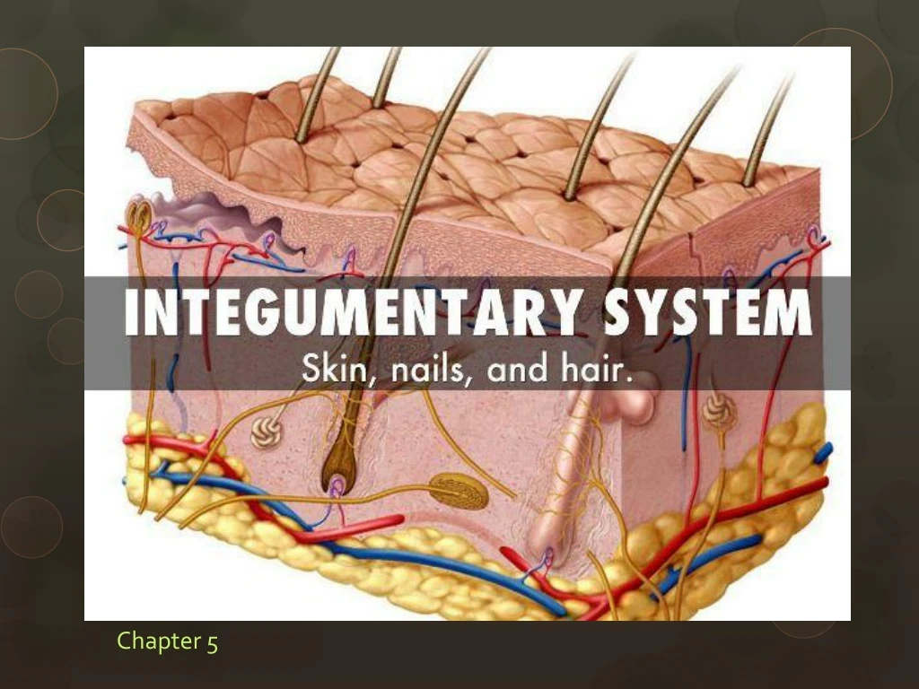
Structure of the skin • Inte- whole • Gument – body • Made up of Epithelial and Connective Tissues • Is the largest and thinnest organ in the body • appendages: hair, nails, glands • one square inch of skin contains more structures than some entire organs • thick skin produces prints and has no hair • cutaneous membrane referred to as the integument
cutaneous membrane referred to as the integument • Cutaneous Membrane : covers the external body surfaces • Adult skin is about 17-20 sq. ft and 10-11 lbs. • thick skin produces prints and has no hair • Ex. Eyelids 1/50” thickness • Ex. Heels 1/8” thickness
Structure of the skin • Layers of the skin: • epidermis: outermost, made of a thin sheet of stratified squamous epithelium • Epi: above • Has hair • dermis: deepest layer, thicker, made mostly of connective tissue • Lacks hair • subcutaneous layer (hypodermis): lies beneath the dermis • made mostly of loose areolar and adipose tissue • insulates, stores energy, shock absorber • Injections are quick and relatively pain free vs. muscle injections • Fat: up to 10 cm in obese individuals
Epidermis • Contain keratin: a tough fibrous protein • strata = layers • melanocytes: cells that produce pigment • cells of the epidermis are held together by desmosomes (cell junctions) • Most of the body is covered by thin skin • Ex. Back, arms, etc (hairy skin) • Thick skin • Palms and feet (no hair)
Cell layers of the epidermis • 1. Stratum corneum; outer most layer of skin • cells continually flake off • Average person sheds about 40 lbs of dead skin cells during their life • 25-30 dead cell layers (flat keratinocytes) • millions of epithelial cells reproduce daily to replace the ones shed • Contains keratin and is our bodies 1st line of defense • 2. Stratum lucidum: clear layer • found in thick skin • Gel substance “eleidin” that will turn into keratin
Cell layers of the epidermis • 3. Stratum granulosum: keratinization begins • Process of cells moving layers – gain keratin as they move up! • Cells begin to regenerate • 4. Stratum spinosum: • Spiny layer (contains RNA) • 5. Stratum basale: base layer (single layer of columnar cells) • only layer where mitosis occurs • new cells move toward surface • Contains keratinocytes (Keratin cells and some stem cells produce new keratinocytes • Stratum Spinosum and Stratum basale make up the growth layer
Epidermal growth and repair • Takes about 35 days to regenerate skin • About 5 weeks • this process will speed up if skin is damaged • Constantly replacing itself all of the time • calluses form when mitotic activity increases due to friction • Causes a thick stratum corneum
Dermis • Deeper of the two layers and much thicker • Called the “true skin” • contains a specialized network of nerves to process sensory info: pain, pressure, touch, and temp • Also includes sweat, oil (subcutaneous gland), blood vessels, and hair follicles.
Layers of the dermis • 1. Papillary layer: made of collagenous and elastic fibers • when elastic fibers decrease wrinkles develop • dermal papillae: parallel rows of projections • binds skin layers together • form ridges and grooves for prints • ridges develop before birth and never change • allow us to walk upright and grip things • Ridges produce friction which give us traction
Layers of the dermis • 2. Reticular layer: dense irregular Connective Tissue with collagen & Elastic Fibers • Serves as part of attachments for many muscles • arrector pili muscles: muscle that contracts only when you are frightened or cold (goosebumps)
Dermal growth and repair • Doesn’t continually regenerate as epidermis does, only during the healing process • Langer’s lines (cleavage lines): patterns formed by the white collagenous fibers • incisions made parallel to cleavage lines will heal better • elastic fibers that are stretched will weaken and tear (stretch marks) • Pregnancy, growth, and weight • Bluish furrows with jagged edges • Heal and lose color (white) • Scars = Dense mass of Connective Tissue • Blisters – separation in epidermis and dermis layers
Skin color • Melanin: determines skin color • number of melanocytes is the same, but the amount of pigment produced varies from person to person • Melanocytes = melanin producing cells • The hormone tyrosinase regulates the release of melanin • Albinism – enzyme tyrosinase is absent at birth
Factors that determine Skin color • Heredity • Inherited from parents • Sunlight – prolonged exposure increase melanin • People that live near the equator • Hormones produced by the pituitary gland • ACTH and MSH • Age • Lower tyrosinase activity = gray hair • carotene: yellow pigment that also contributes to skin color
Skin color • Skin can change color depending on blood flow and oxygen • Hemoglobin • Oxygen carrying pigment in RBCs • cyanosis: decrease in blood flow causes the skin to turn bluish gray • Not enough oxygen in the blood • Vessels constrict = pale • Vessels dilate = pinker


