CHROMOSOME VARIATION
CHROMOSOME VARIATION. VARIATION IN CHROMOSOME NUMBER. Variations in Chromosome Number.

CHROMOSOME VARIATION
E N D
Presentation Transcript
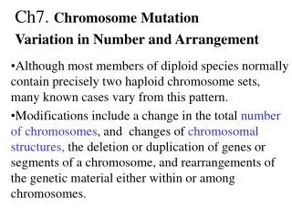
VARIATION IN CHROMOSOME NUMBER
Variations in Chromosome Number An organism or cell is euploid when it has one complete set of chromosomes, or exact multiples of complete sets. Eukaryotes that are normally haploid or diploid are euploid, as are organisms with variable numbers of chromosome sets. Aneuploidy results from variations in the number of individual chromosomes (not sets), so that the chromosome number is not an exact multiple of the haploid set of chromosomes.
Changes in Complete Sets of Chromosomes Monoploidy and polyploidy involve complete sets of chromosomes, and so both are cases of euploidy. Euploidy is lethal in most animal species, but often tolerated in plants, where it has played a role in speciation and diversification.
Monoploidy and polyploidy can result when either round of meiotic division lacks cytokinesis, or when meiotic nondisjunction occurs for all chromosomes. • Complete nondisjunction at meiosis II will produce 1⁄2 gametes with normal chromosomes, 1⁄4 with two sets of chromosomes and 1⁄4 with no chromosomes. • A gamete with two sets of chromosomes fused with a normal gamete produces a triploid (3N) zygote. • Fusion of two gametes that each have two sets of chromosomes produces a tetraploid (4N) zygote. • Polyploidy of somatic cells can result from mitotic nondisjunction of complete chromosome sets.
Monoploidy is rare in adults of diploid species due to recessive lethal mutations. • Males of some species (e.g., wasps, ants and bees) develop from unfertilized eggs and are monoploid.
Plant experiments often use monoploids. • Haploid cells are isolated from plant anthers and grown into monoploid cultures. • Colchicine (which inhibits mitotic spindle formation) allows chromosome number to double, producing completely homozygous diploid breeding lines. • Mutant genes are easily identified in monoploid organisms.
Polyploidy involves three or more sets of chromosomes, and may occur naturally (e.g., by breakdown of the mitotic spindle), or by induction (e.g., with chemicals such as colchicine). • Nearly all plants and animals probably have some polyploid tissues. Examples: • Plant endosperm is triploid. • Liver of mammals (and perhaps other vertebrates) is polyploid. • Wheat is hexaploid (6N) and the strawberry is octaploid (8N). • North American sucker fish, salmon and some salamanders are polyploid.
There are two classes of polyploids based on the number of chromosome sets: • Even-number polyploids are more likely to be at least partially fertile, because the potential exists for equal segregation of homologs during meiosis. • Odd-number polyploids will always have unpaired chromosomes. Balanced gametes are rare and these organisms are usually sterile or have increased zygote death.
Triploids are unstable in meiosis, because random segregation means that balanced gametes (either exactly N or exactly 2N) are rare. • The probability of a triploid organism producing a haploid gamete is (1⁄2)n, where n is the number of chromosomes. • Triploidy is always lethal in humans, accounting for 15–20% of spontaneous abortions and 1/104 live births, with most dying in the first month. • Tetraploidy in humans is also lethal, usually before birth, accounting for 5% of spontaneous abortions.
Triploids in plants can be valuable in plants because they are “seedless”. Examples are bananas, watermelons.
Polyploidy is more common in plants, probably due to self-fertilization, allowing an even-number polyploids to produce fertile gametes and reproduce. Plant polyploidy occurs in two types: Autopolyploidy results when all sets of chromosomes are from the same species, usually due to meiotic error.
Allopolyploidy results when the chromosomes are from two different organisms, typically from the fusion of haploid gametes followed by chromosome doubling. Fusion of haploid gametes from plant 1 and plant 2 produces an N1 + N2 hybrid plant. No chromosomal pairing occurs at meiosis, viable gametes are not produced and the plants are sterile.
Rarely, division error doubles the chromosome sets (2 N1 + 2N2). The diploid sets function normally in meiosis, and fertile allotetraploid plants result. Polyploidy is the rule in agriculture, where polyploids include all commercial grains (e.g., bread wheat, Triticum aestivum, an allohexaploid of three plant species), most crops and common flowers.
Changes in One or a Few Chromosomes Aneuploidy vs Euploidy
Aneuploidy can occur due to nondisjunction during meiosis. Nondisjunction during meiosis I will produce four gametes, two with a chromosome duplicated, and two that are missing that chromosome. • Fusion of a normal gamete with one containing a chromosomal duplication will produce a zygote with three copies of that chromosome, and two of all others. • Fusion of a normal gamete with one missing a chromosome will result in a zygote with only one copy of that chromosome, and two of all others.
Nondisjunction during meiosis II produces two normal gametes and two that are abnormal (one with two sibling chromosomes, and one with that chromosome missing). Fusion of abnormal gametes with normal ones will produce the genotypes discussed above. Normal gametes are also produced, and when fertilized will produce normal zygotes.
Autosomal aneuploidy is not well tolerated in animals, and in mammals is detected mainly after spontaneous abortion. Aneuploidy is much better tolerated in plants. There are four main types of aneuploidy: • Nullisomy involves loss of 1 homologous chromosome pair (the cell is 2N - 2). • Monosomy involves loss of a single chromosome (2N - 1). • Trisomy involves one extra chromosome, so the cell has three copies of one, and two of all the others (2N + 1). • Tetrasomy involves an extra chromosome pair, so the cell has four copies of one, and two of all the others (2N + 2).
More than one chromosome or chromosome pair may be lost or added. Examples: • A double monosomic aneuploidy has two separate chromosomes present in only one copy each (2N - 1 - 1). • A double tetrasomic aneuploidy has two chromosomes present in four copies each (2N + 2 + 2).
Some types of aneuploidy have serious meiotic consequences. Examples: • A monosomic cell (2N - 1): • May produce gametes that are N (normal) and N - 1(monosomic). • Or, the unpaired chromosome may be lost completely, producing gametes that are all N - 1.
Trisomics • Important genetic tools to understand dosage effects • AAA, AAa, Aaa, aaa • Used for linkage analysis in plants
A trisomic cell (2N + 1) with the genotype +/+/a (assuming that this organism can tolerate trisomy, and no crossing-over occurs). • Gametes produced belong to four genotypic classes, in these proportions: (1) Two gametes with genotype +/a. (2) Two gametes with genotype +. (3) One gamete with genotype +/+. (4) One gamete with genotype a.
The cross of a +/+/a trisomic to an a/a individual will produce a phenotypic ratio of 5 wild type : 1 mutant (a). Really just look for altered segregation numbers.
VARIATIONS IN CHROMOSOME STRUCTURE (CHROMOSOMAL REARRANGEMENTS)
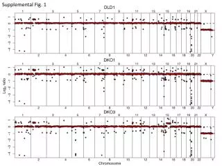
Genomic DNA Variation = variations between the genome of different individuals • Single Nucleotide Polymorphisms (SNPs) = substitutions, short indels (one to a few nucleotides) • Micro- and mini-satellite expansion and contraction (typically less than 100 bp variation) • Satellite DNA expansion and contraction (unit >100 bp, mostly centromeric) • Transposable Elements insertion/excision (ranging from ~100 bp to less than 10 kb) • Segmental Duplications = Low copy repeats (LCRs) (>1 kb- 3 Mb with similarity >90%) -- include copy number variants (CNVs) (submicroscopic) • Large chromosomal rearrangements: Mb-range duplication, insertion, deletion (CNVs), inversion, translocation (microscopic structural variation) • Changes in chromosome numbers = aneuploidy (typically deleterious) (microscopic structural variation)
Types of Chromosomal Mutations Variations in chromosome structure or number can arise spontaneously or be induced by chemicals or radiation. Chromosomal mutation can be detected by: • Genetic analysis (observing changes in linkage). • Microscopic examination of eukaryotic chromosomes at mitosis and meiosis (karyotype analysis).
Types of Chromosomal Mutations Chromosomal aberrations contribute significantly to human miscarriages, stillbirths and genetic disorders. • About 1⁄2 of spontaneous abortions result from major chromosomal mutations. • Visible chromosomal mutations occur in about 6/1,000 live births. • About 11% of men with fertility problems, and 6% of those institutionalized with mental deficiencies have chromosomal mutations.
Variations in Chromosome Structure Mutations involving changes in chromosome structure occur in four common types: a. Deletions. b. Duplications. c. Inversions (changing orientation of a DNA segment). • Translocations (moving a DNA segment). All chromosome structure mutations begin with a break in the DNA, leaving ends that are not protected by telomeres, but are “sticky” and may adhere to other broken ends.
Chromosomal translocation revealed by ‘chromosome painting’ (or spectral karyotyping)
Four major types: • Deletions • Duplications • Inversions • Translocations
Deletion In a deletion, part of a chromosome is missing. Deletions start with chromosomal breaks induced by: • Heat or radiation (especially ionizing). • Viruses. • Chemicals. • Transposable elements. • Errors in recombination. Deletions do not revert, because the DNA is missing.
Deletion The effect of a deletion depends on what was deleted. • A deletion in one allele of a homozygous wild-type organism may give a normal phenotype, while the same deletion in the wild-type allele of a heterozygote would produce a mutant phenotype. • Deletion of the centromere results in an acentric chromosome that is lost, usually with serious or lethal consequences. (No known living human has an entire autosome deleted from the genome.) • Large deletions can be detected by unpaired loops seen in karyotype analysis.