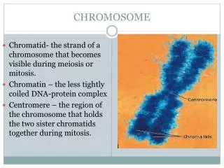
CHROMOSOME
Chromatid- the strand of a chromosome that becomes visible during meiosis or mitosis. Chromatin – the less tightly coiled DNA-protein complex Centromere – the region of the chromosome that holds the two sister chromatids together during mitosis. CHROMOSOME.

CHROMOSOME
E N D
Presentation Transcript
Chromatid- the strand of a chromosome that becomes visible during meiosis or mitosis. Chromatin – the less tightly coiled DNA-protein complex Centromere – the region of the chromosome that holds the two sister chromatids together during mitosis. CHROMOSOME
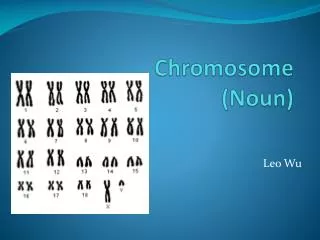
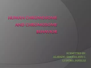
Sex chromosomes are chromosomes that determine the sex of an organism, and they may also carry genes for other characteristics. In humans, sex chromosomes are either X or Y. XX for female XY for male The autosomes are all the other chromosomes in an organism except for sex chromosomes. In humans two of the 46 are sex chromosomes, and the remaining 44 chromosomes are autosomes. SEX CHROMOSOMES & AUTOSOMES
The DNA in eukaryotic cells wraps tightly around proteins called histones. Histones help maintain the shape of the chromosomes and aid in the tight packaging of DNA. Nonhistone proteins are generally involved in controlling the activity of specific regions of the DNA HISTONE AND NON-HISTONE
CHROMOSOME Each chromosome contains a constricted region called the centromere, whose location establishes the general appearance of each chromosome chromosomes are classified as metacentric, submetacentric, acrocentric, or telocentricon the basis of the centromere location The shorter arm, by convention, is shown above the centromere and is called the p arm (p, for “petite”). The longer arm is shown below the centromere and is called the q arm (q because it is the next letter in the alphabet).
CELL CYCLE The cell cycle is an orderly set of stages that take place between the time a cell divides and the time the resulting daughter cells also divide.
Interphase -the cell carries on its regular activities. The three stages: G1 phase S phase G2 phase. Mitotic Stage Stages of mitosis: karyokinesis (division of the nucleus) cytokinesis (division of the cytoplasm). CELL CYCLE STAGES
Interphase and the Cell Cycle The events that occur from the completion of one division until the completion of the next division constitute the cell cycle. Interphase: G1 phase, S-phase & G2 phase
Mitosis Partitions Chromosomesinto Dividing Cells PURPOSE OF MITOSIS In some single-celled organisms, such as protozoans and some fungi and algae, mitosis provides the basis for asexual reproduction. foundation for the development and growth of the organism (zygote). In adult multicellular organism, mitotic activity is the basis for wound healing and other forms of cell replacement in certain tissues.
MITOSIS KARYOKINESIS CYTOKINESIS This process is quite complex and requires great precision. The chromosomes must first be exactly replicated and then accurately partitioned. The end result is the production of two daughter nuclei, each with a chromosome composition identical to that of the parent cell. This less complex process requires a mechanism that partitions the volume into two parts, then encloses each new cell in a distinct plasma membrane. As the cytoplasm is reconstituted, organelles either replicate themselves, arise from existing membrane structures, or are synthesized de novo (anew) in each cell.
PROPHASE MAJOR EVENTS In animal cells, migration of two pairs of centrioles to opposite ends of the cell (plant cells do not have cemtrioles). the nuclear envelope begins to break down and gradually disappears. the nucleolus disintegrates within the nucleus the diffuse chromatin fibers have begun to condense, until distinct threadlike structures, the chromosomes, become visible.
PROPHASE MAJOR EVENTS (cont) Near the end of prophase the chromosomes become apparent and is actually a double structure split longitudinally except at a single point constriction, the centromere. The two parts of each chromosome are called sister chromatids
PROMETAPHASE and METAPHASE Prometaphaserefers to the period of chromosome movement and the term metaphase is applied strictly to the chromosome configuration following migration
Metaphase During metaphase, the kinetochore fibers move the chromosmes to the center of the dividing cell. Once the chromosomes are in the center of the cell, each of them is held in place by the kinetochore fibers. .
Anaphase Major Events For complete disjunction: During this phase, sister chromatids of each chromosome, held together only at their centromere regions, disjoin (separate) from one another—an event described as disjunction—andare pulled to opposite ends of the cell. (1), shugoshin must be degraded, reversing its protective role; (2), the cohesin complex holding the centromere region of each sister chromosome is then cleaved by separase; and (3) sister chromatids of each chromosome are pulled toward the opposite poles of the cell
Telophase Major Events At its beginning, two complete sets of chromosomes are present, one set at each pole. The most significant event of this stage is cytokinesis, the division or partitioning of the cytoplasm Animal cells and plant cells differ in cytokinesis
Plant and Animal Cells’ Cytokinesis Plants Animals In plant cells, a cell plate is synthesized and laid down across the region of the metaphase plate. Animal cells, however undergo a constriction of the cytoplasm, much as a loop of string might be tightened around the middle of a balloon.
Telophase Major events (cont) In each new cell, the chromosomes begin to uncoil and become diffuse chromatin once again, while the nuclear envelope reforms around them, the spindle fibers disappear, and the nucleolus gradually reforms and becomes visible in the nucleus during early interphase.
Again. . . What are the roles of the following during cell division? Cohesin (in cell division) Shugoshin Microtubule Separase
CONTROL OF CELL DIVISION In eukaryotes, proteins regulate the progress of cell division at certain checkpoints.
1. Cell Growth Checkpoint. Proteins at this checkpoint control whether the cell will divide. Hint: if the cell is healthy and has grown to a suitable size during G1 phase, protein will initiate DNA synthesis (S phase). If conditions are not favorable for DNA synthesis, the cell cycle will stop at this point. CELL GROWTH CHECKPOINT
2. DNA Synthesis (G2) checkpoint. DNA repair enzymes check the results of DNA replication. If this checkpoint is passed, proteins will signal the cell to begin the molecular processes that will allow the cell to divide mitotically. DNA SYNTHESIS CHECKPOINT
3. Mitosis checkpoint. If a cell passes this checkpoint, proteins signal the cell to exit mitosis. The cell enters into the G1 phase, the major growth phase of the cell cycle, once again. MITOSIS CHECKPOINT
PROKARYOTIC CELL DIVISION Most prokaryotes reproduce by binary fission, in which two identical cells are produced from one cell.
Meiosis Reduces the ChromosomeNumber from Diploid to Haploidin Germ Cells and Spores
Three (3) Characterized Events in Prophase I First First, as in mitosis, chromatin present in interphase thickens and coils into visible chromosomes. And, as in mitosis, each chromosome is a double structure, held together by the molecular complex called cohesin. . Second, unlike mitosis, members of each homologous pair of chromosomes pair up, undergoing synapsis. Third Crossing over occurs between chromatids of synapsedhomologs
PROPHASE 1 - LEPTONEMA Leptotene Stage the interphasechromatin material begins to condense, and the chromosomes, though still extended, become visible. Along each chromosome are chromomeres, localized condensations that resemble beads on a string. homology search, which precedes and is essential to the initial pairing of homologs.
PROPHASE I - ZYGONEMA Zygotene Stage The chromosomes continue to shorten and thicken. During the process of homology search, homologous chromosomes undergo initial alignment with one another - rough pairing is complete by the end of zygonema. structures called lateral elements are visible between paired homologs. synaptonemal complex begins to form between the homologs (for pairing of homologs) It is upon completion of zygonema that the paired homologs are referred to as bivalents.
PROPHASE I - PACHYNEMA Pachytene Stage the chromosomes continue to coil and shorten, further development of the synaptonemal complex between bivalent for more intimate pairing. each homolog is now evident as a double structure, providing visual evidence of the earlier replication of the DNA of each chromosome (bivalent contains 4-member chromatids)
PROPHASE I - DIPLONEMA DIPLOTENE STAGE more apparent that each tetrad consists of two pairs of sister chromatids. Within each tetrad, each pair of sister chromatids begins to separate. However, one or more areas remain in contact where chromatids are intertwined. – (chiasma is an area for crossing-over)
PROPHASE I - DIAKINESIS during this final substage, the nucleolus and nuclear envelope break down the two centromeres of each tetrad attach to the recently formed spindle fibers The chromosomes pull farther apart, but nonsisterchromatids remain loosely associated at the chiasmata. As separation proceeds, the chiasmata move toward the ends of the tetrad.
Metaphase, Anaphase, and Telophase I metaphase I Anaphase I the chromosomes have maximally shortened and thickened. The terminal chiasmata of each tetrad are visible and appear to be the major factor holding the nonsisterchromatids together. Each tetrad interacts with spindle fibers, facilitating its movement to the metaphase plate. cohesin is degraded between sister chromatids, except at the centromere region, which, as in mitosis, is protected by a shugoshin complex. Then, one-half of each tetrad (a dyad) is pulled toward each pole of the dividing cell.
Telophase I reveals a nuclear membrane forming around the dyads and the cells go directly to meiosis II without chromosome replication. In general, meiotic telophase is much shorter than the corresponding stage in mitosis.
The Second Meiotic Division MEIOSIS II essential if each gamete or spore is to receive only one chromatid from each original tetrad
MEIOSIS II PROPHASE II METAPHASE II Each dyad is composed of one pair of sister chromatids attached by the common centromeric region. the centromeres are positioned on the equatorial plate. the shugoshin complex is degraded and separates the centromeres
Anaphase II Telophase II the sister chromatids of each dyad are pulled to opposite poles. onemember of each pair of homologous chromosomes present at each pole. each chromosome is now a monad four haploid gametes may result from a single meiotic event