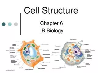
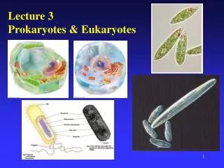
Lecture 3 Prokaryotes & Eukaryotes
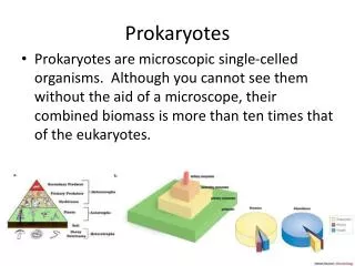
Lecture 3 Prokaryotes & Eukaryotes. Components of ALL cells Plasma membrane = lipid bilayer that forms a physical barrier to all cells Cytoplasm = the semisolid components within the cell (Cytosol = Fluid portion) Chromosomes = DNA structure containing genes

Lecture 3 Prokaryotes & Eukaryotes
E N D
Presentation Transcript
Lecture 3 Prokaryotes & Eukaryotes
Components of ALL cells • Plasma membrane= lipid bilayer that forms • a physical barrier to all cells • Cytoplasm= the semisolid components within the cell (Cytosol = Fluid portion) • Chromosomes= DNA structure containing genes • Ribosomes = tiny structures of RNA/protein • that synthesize new proteins using instructions from the genes
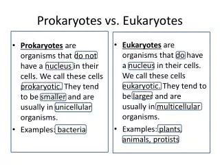
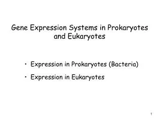
Prokaryotes v. Eukaryotes nucleoid versus a nucleus Cell size - Prokaryotic cells are smaller (Most 0.2 - 5 µm) 1000 X magnification - Eukaryotic cells are larger (Most 10 - 100 µm) 200 to 500 X magnification Membrane-bound organelles - Almost none in prokaryotes - Eukaryotic cells have many organelles of specialized form and function - Complex cytoskeleton composed of various types of filaments - Large ribosomes
Distinguishing Features of Prokaryotes • Nucleoid • No histones • No numerous organelles • Cell walls • Peptidoglycan • Binary fission • Pili or fimbriae • Single Circular Chromosome • Some exceptions • Plasmids • Smaller usually circular pieces of DNA
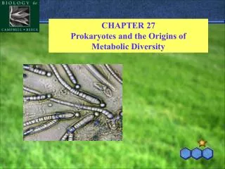
The Prokaryotic Cell Wall Many Types of Bacterial Cell Walls but Two Main Types… 1. Gram Positive 2. Gram Negative Both contain peptidoglycan but differ on amount
Hans Christian Gram (1853-1938) Hans Christian Gram was a Danish doctor studying in Berlin who studied lung tissues of pneumonia victims. He noticed that different bacteria behaved differently when stained with a cationic dye and classified them as Gram positive (stained) or Gram negative (didn’t stain).
Gram Stain Slide Gram + have a thick layer of peptidoglycan
N-acetylmuramic acid (NAM) Peptidoglycan • Macromolecule composed of a repeating framework of long chains cross-linked by short peptide fragments • Unique to bacteria • Composed of 2 sugars: NAG & NAM • Sugars alternate in the backbone • Rows linked by polypeptides • Provides strong, flexible support to keep bacteria from bursting or collapsing because of changes in osmotic pressure N-acetylglucosamine (NAG)
Be able to identify all the parts of a Gram + & - cell wall for the next exam.
Gram-positive versus Gram-negative Cell Walls Fig. 4.16 Thick – 20-80 nm Thin – 8-11 nm Teichoic acids are found only in Gram-positive cell walls. They are negatively charged and their function is unknown. Lipopolysaccharide and outer membrane are only found in Gram-negative cell walls.
Eukaryotic Cell Wall • Animal cells have no cell wall • Elaborate extracellular matrices • Collagen & glycoproteins • No eukaryotic cell has peptidoglycan in their cell wall • Peptidoglycan is unique to bacteria • Many eukaryotes have a cell wall composed of a carbohydrate • Cell wall of algae & plants is made of cellulose • A carbohydrate chain • Cell wall of fungi is made of chitin • A carbohydrate chain • Cell wall of yeasts is made of glucan and mannan • A carbohydrate chain
Plant Cell Wall Cellulose
Fungal cell Illustration shows relationship between the cell membrane and cell wall. Glycocalyx is the outermost section.
All Cells have a Membrane Plasma membrane functions as a selective barrier O2 & nutrients must enter the cell Waste products must exit the cell
Lipid Bilayer • Two layers of phospholipids • Main component of cell membranes • Membrane has fluid properties • Most phospholipids and some proteins can drift through membrane • It’s FLUID & not static
Fluid Mosaic Model • Membrane is a mosaic of • Phospholipids • Glycolipids Carbohydrates • Sterols • Eukaryotes • Proteins
Prokaryotes Lipid bilayer Selectively permeable Allows secretion Site for metabolic rxns Respiration Photosynthesis Nutrient processing Synthesis of proteins & other molecules Eukaryotes Lipid bilayer Selectively permeable Endocytosis Exocytosis Sterols Cholesterol Reinforces cell wall All organelles have a membrane very similar to the cell membrane Cell Membrane Pro v. Euk
Cell membranes of both Prokaryotes and Eukaryotes perform • Diffusion, osmosis & active transport • Endocytosis is unique to Eukaryotes • Phagocytosis • Uses pseudopods - surround and engulf • Pinocytosis • Cell drinking • Plasma membrane folds in on itself • Often times receptor mediated • Exocytosis
Cell Membranes Show Selective Permeability Other Mechanisms Diffusion
Plasma Membrane • Passive Transport • Diffusion • Active Transport • Requires energy as ATP (adenosine triphosphate) • Cell membrane proteins carry out many tasks • Highly specialized proteins • Enzymes • Recognition and signaling • Energy Reactions (Prokaryotes)
Osmosis • Diffusion of water molecules across a selectively permeable membrane • Direction of net flow is determined by water concentration gradient • Side with the most solute molecules has the lowest water concentration
Biological Relevance of Osmosis Cells can be in one of 3 conditions Plasmolysis = death Cell lysis = death Isotonic - solutes balanced Hypotonic - more solute inside cell Hypertonic - more solute outside cell Good Situation!
Animal Cell [H2O] is greater in the cell than outside [H2O] is greater outside the cell than inside
Plant Cell Hypertonic Hypotonic Isotonic [H2O] is greater in the cell than outside [H2O] is greater outside the cell than inside
Flagella & Cillia Prokaryotic & Eukaryotic flagella are not similar in size or structure
Prokaryotic Flagella • Long filamentous projections used to propel bacteria – 18-20 nm • Several types of flagella arrangements Monotrichous – One flagellum usually at one pole (Polar) Amphitrichous - tufts of flagella at both poles Lophotrichous - two or more flagella at one pole Petritrichous - flagella distributed over entire surface
Parts of the Flagellum • Filament - main body of • flagella, made of flagellin • Hook - attaches flagella to basal • body • Basal body - attaches flagella to • cell • Movement is accomplished by • rotating basal body • ATP • Results in rotation of filament • Smooth running or tumbling
Direction of Flagellar Rotation Important for Motility • Counterclockwise rotation • results in movement • Clockwise results in tumbling • Responds to chemo and • phototaxis possible • Attractants induce “running” • Repellants induce “tumbling” • This model is for Escherichia coli
Axial Filaments • Spirochetes • Endoflagella • Bundle of fibrils • Run length of organism • Drives spirochete forward • in a spiral motion
Eukaryotic Flagella and Cilia • Flagella - long (~40 um), few • 10X larger (diameter) than Pro flagella (~180-200 nm in width) • Cilia – short (10 um long), many • Oars • Ciliated protozoa & animal cells • Both used for motility • Both have 9+2 microtubule structure • Hollow tubes that slide past one another (made up of tubulin) • Waves and whips • Doesn't rotate - different from bacterial flagella • Pull & push
Motor Protein Dynein ATP
Prokaryotic Genome Size Bacteria Size (Mbp) Escherichia coli 4.64 Bacillus subtilis 4.20 Streptococcus pyrogenes 1.85 Mycobacterium genitalium 0.58 Archaea Size (Mbp) Methanococcus jannaschii 1.66 Sulfolobus solfactaricus 2.25 Pyrococcus furiosus 1.75 Genome consists of usually one circular chromosome and plasmids (if present)
Eukaryotic Genome Size Organism Mbp Homo sapiens 3000 Drosophilia melanogaster 165 Plasmodium falciparum 23 Saccharomyces cerevisiae 12.07 Eukaryotes also have Mitochondrial DNA Chloroplast DNA Genome usually consists of a number of linear chromosomes
Glycocalyx, Capsule & Slime Capsule Repeating units of polysaccharide, protein or both (a polymer) Adheres tightly, thick & gummy Mostly a Prokaryotic term – interchangeable with glycocalyx Slime Layer Polysaccharide, protein or both that is easily washed off Mostly a Prokaryotic term Glycocalyx Outermost layer of cell that come into contact with environment This term is used for both Eukaryotes and Prokaryotes Sticky carbohydrates attached to proteins Important in protection & adhesion
Pili & Fibriae • Prok surface appendages • Pilus is longer • Gram negative bacteria • Conjugation • Fibria is shorter • Bristlelike • Stick to surfaces • Colonize host tissue • Euk do not produce these structures
Ribosomes – RNA and Protein Prok Euk All ribosomes are made up of two subunits
Prok Ribosomes Smaller 70S 50S & 30S Free ribosomes Located in the cytosol Euk Ribosomes Larger 80S 60S & 40S Free ribosomes Located suspended in the cytosol Synthesize proteins that function within the cytosol Bound ribosomes Are attached to the outside of the endoplasmic reticulum Synthesize proteins that are included into membranes or exported from the cell
Organelles • Membrane-bounded functional units • Compartmentalized tasks instead of a mixture • Allows for much more variety of functionality • Present in Euk • Absent in Prok • Prok conduct the similar activities at the cell membrane
A View of the Eukaryotic Cell Much more complex - many levels of compartmentalization
The Nucleus • Largest organelle - contains DNA • Enclosed by double layered lipid envelope • Pores allow transport of various cytoplasmic substances • Contain nucleoli - sites of rRNA synthesis • DNA organized by histones • Further organized into chromatin - thread like • Further condenses to chromosomes for replication
The Endoplasmic Reticulum • Extensive network of flattened cisterns continuous with the nuclear envelope • Rough ER - studded with ribosomes • Protein entry point • Modifications made, lipids and carbohydrates attached • Smooth ER - no ribosomes • More enzymatic diversity • Synthesize lipids, oils, phospholipids, steroids
Be able to identify all structures listed in this illustration
The Golgi Complex • Receives all proteins transported from RER • Mail station of the cell - all proteins sorted for transport • Composed of cisterns - flattened membranous stacks • Many post-translational modifications made • Determines fate of protein • Packaged into secretory • vesicle • Can be packaged into • transport vesicle (transfer • between stacks, transfer to • storage vesicles)