Download
1 / 66
730 likes | 898 Vues
Learn about different types and patterns of traumatic elbow instability, associated fractures, and ligament injuries, including chronic and recurrent dislocations. Discover treatment options and surgical protocols for managing these complex elbow conditions.

E N D
Traumatic Elbow Instability David Ring MD PhD Updated April 2016
Simple Elbow Dislocation • No associated fractures • Complete or near complete capuloligamentous injury • Extensive muscle injury • Nearly always stable after reduction • No advantage to surgery if stable • No more than 2 weeks immobilization
Elbow Dislocation • Usually posterolateral • Can dislocate with anterior band of MCL intact • Posteromedial pattern • Less common • Possibly more unstable
Slight Subluxation • “Drop Sign” • This is like pseudo-subluxation in the shoulder. • The combination of extensive muscle and ligament injury and guarding due to pain create a slight sag. • IMPORTANT: distinguish from subluxation that will cause articular damage • Drop sign • After active flexion exercises
Slight Subluxation • “Drop Sign” • Management: • Avoid varus stress (shoulder abduction) • Active flexion • Overhead exercises • Drop sign • After active flexion exercises
Unstable Simple Elbow Dislocation • Uncommon • Older women (simple fall) • Young men (high-energy)
Unstable Simple Elbow Dislocation • Ligament / muscle reattachment to epicondyles • External fixation • Cross pinning
Cross Pinning • Useful bail out • Stiff and located is preferred to subluxation • Stiffness usually worked out easily • 2.0mm pins exit proximally for retrieval in case of breakage • Can be placed with local • Only needed for 3 weeks. Bury if needed longer
Chronic Simple Elbow Dislocation Jupiter and Ring JBJS 2002 • Treatment: Open reduction and hinged external fixation • No ligament reconstruction • 5 patients: dislocated for 2 to 9 months • Stable elbow, > 100 degrees motion in all patients
Medial Collateral Ligament Insufficiency • Throwing athletes • Chronic attenuation • Inability to throw 95 mph fastballs
LCL Insufficiency Recurrent Simple Elbow Dislocation • Insufficiency of the lateral collateral ligament • Adolescent elbow dislocation • Iatrogenic
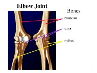
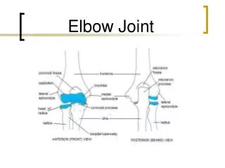
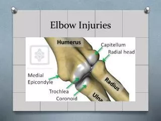
Definition • Fracture-dislocation of the elbow • Dislocation of the elbow • Intra-articular fracture
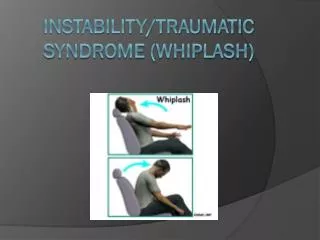
Definition • Traumatic Elbow Instability • Injury that destabilizes the elbow • With or without dislocation
Patterns of Traumatic Elbow Instability With Fracture Dislocation with Articular Fracture • Varus posteromedial rotational instability Olecranon Fracture-Dislocations Dislocation + radial head fracture Anterior Terrible Triad Posterior
Dislocation vs. Disruption • Dislocation • Disruption
Dislocation vs. Disruption • Dislocation • Disruption
Dislocation vs. Disruption • Dislocation • Disruption • Ligaments Partially Spared
Disruption • Ligaments Partially Spared
Patterns of Traumatic Elbow Instability With Fracture • Dislocation Injuries • Disruption Injuries Dislocation with Articular Fracture • Varus posteromedial rotational instability Olecranon Fracture-Dislocations Dislocation + radial head fracture Anterior Terrible Triad Posterior
Posterior Dislocation + Radial Head Fracture • 24 patients • Ulnohumeral dislocation with radial head fracture • Cast 1 month +/- radial head resection • “Results better than generally thought” • Secondary procedures for radial head • No problems with instability
Posterior Dislocation + Radial Head Fracture • 23 patients • Excision of radial head and cast • INSTABILITY in patients with CORONOID fractures (4 patients)
Terrible Triad • Posterior dislocation • Radial head fracture • Coronoid fracture
Terrible Triad • Only patients with INSTABILITY had CORONOID fractures (4 patients)
Terrible Triad Ring, Jupiter, Zilberfarb JBJS 2002 • 11 patients • Regan and Morrey Type 2 coronoid fractures • 7 redislocated in splint or cast • 5 redislocated after operation • Only 4 patients with satisfactory results
Terrible Triad Pugh DM, Wild LM, Schemitsch EH, King GJ, McKee MD • Standard surgical protocol to treat elbow dislocations with radial head and coronoid fractures. • J Bone Joint Surg Am. 2004 Jun;86-A(6):1122-30.
Regan and Morrey • Based on single lateral radiograph • Type 1: Tip avulsion • Type 2: < 50% coronoid height • Type 3: > 50% coronoid height
O’Driscoll Classification • 3 • 1 • 2
Varus Posteromedial Rotational Injuries • Inadequate Treatment
Olecranon Fracture-Dislocations • Anterior (trans-olecranon) fracture-dislocations • Posterior (posterior Monteggia) fracture-dislocations
Anterior (Trans-Olecranon Fracture-Dislocation of the Olecranon
Anterior (Trans-Olecranon) Fracture-Dislocation of the Olecranon
Posterior Fracture-Dislocation of the Olecranon POSTERIOR MONTEGGIA TYPE FRACTURE-DISLOCATION
Posterior Fracture-Dislocation of the Olecranon POSTERIOR MONTEGGIA TYPE FRACTURE-DISLOCATION
Principles of Treatment • Restore contour and dimensions of trochlear notch • Contoured dorsal plate • Fixation of coronoid • Bridge fragmentation
Treatment Tips • Pin the olecranon to the trochlea • Consider a temporary external fixator for a complex fracture
Coronoid Exposure • Through an olecranon fracture • Lateral • Kaplan interval with elevation of ECRL origin • Removal of radial head fragments • Medial • Over the top (tip) • Split in FCU by ulnar nerve (medial facet) • Elevate entire flexor-pronator mass from dorsal (base fracture)
Coronoid Provisional Fixation • Coronoid fixation with plate then reduce and fix olecranon • Pin fragments to trochlea • Need to immobilize the elbow