Nerve-Muscle Interaction
Nerve-Muscle Interaction. Skeletal muscle activation is initiated through neural activation NS can be divided into central (CNS) and peripheral (PNS) The NS can be divided in terms of function: motor and sensory activity

Nerve-Muscle Interaction
E N D
Presentation Transcript
Nerve-Muscle Interaction • Skeletal muscle activation is initiated through neural activation • NS can be divided into central (CNS) and peripheral (PNS) • The NS can be divided in terms of function: motor and sensory activity • Sensory: collects info from the various sensors located throughout the body and transmits the info to the brain • Motor: conducts signals to activate muscle contraction
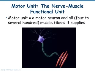
Activation of motor unit and its innervation systems • Spinal cord 2. Cytosome 3. Spinal nerve • 4. Motor nerve 5. Sensory nerve 6. Muscle with muscle fibres
Motor Unit • Motor nerves extend from the spinal cord to the muscle fibres • Each fibre is activated through impulses delivered via motor endplate • Motorunit: a group of fibres activated via the same nerve • All muscle fibres of one particular motor unit are always of the same fibre type • Muscles needed to perform precisemovements generally consist of a large number of motor units and few muscle fibres • Lessprecisemovements are carried out by muscles composed of fewer motor units with many fibres per unit
All-or-none Principle • Whether or not a motor unit activates upon the arrival of an impulse depends upon the so called all-or-none principle • An impulse of a certain magnitude (or strength) is required to cause the innervated fibres to contract • Every motor unit has a specific threshold that must be reached for such activation to occur
Intra-muscle Coordination • The capacity to apply motor units simultaneously is known as intra-muscle coordination • Many highly trained power athletes, such as weightlifters, wrestlers, and shot putters, are able to activate up to 85% of their available muscle fibres simultaneously (untrained: 60%) • Force deficit: the difference between assisted and voluntarily generated maximal force (trained: 10%, untrained: 20-35%)
Intra-muscle Coordination cont. • Trained athletes have not only a larger muscle mass than untrained individuals, but can also exploit a larger number of muscle fibres • Athletes are more restricted in further developing strength by improving intra-muscular coordination • Trained individuals can further increase strength only by increasing muscle diameter
Inter-muscle Coordination • The interplay between muscles that generate movement through contraction (agonists) and muscles responsible for opposing movement (antagonists) is called inter-muscle coordination • The greater the participation of muscles and muscle groups, the higher the importance of inter-muscle coordination • To benefit from strength training the individual muscle groups can be trained in relative isolation • Difficulties may occur if the athlete fails to develop all the relevant muscles in a balanced manner
Inter-muscle Coordination cont. • High-level inter-muscle coordination greatly improves strength performance and also enhances the flow, rhythm, and precision of movement • Trained athlete is able to translate strength potential to enhance inter-muscle coordination
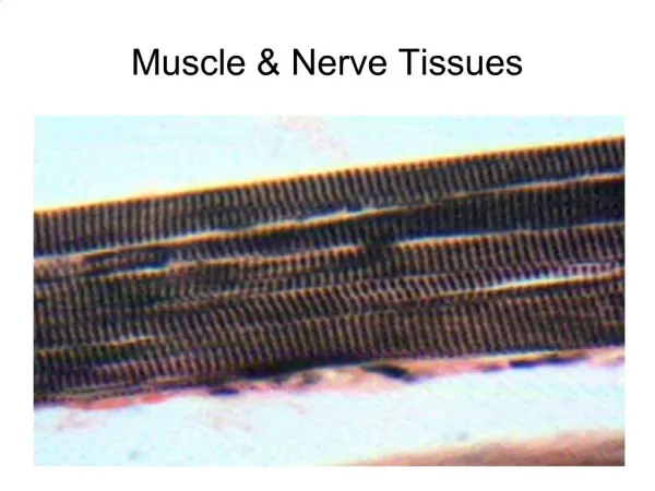
Sarcomeres • separated by narrow zones of dense material called Z lines • within a sarcomere is a dark area called the A band (thick myofilaments) • ends of the A band are darker because of overlapping thick and thin myofilaments • the light coloured area is called the I band (thin myofilaments) • the combination of alternating dark A bands and light I bands gives the muscle fibre its striated appearance
Muscle Contraction • Muscle structure under a microscope Muscle fibres • skeletal muscle viewed under a microscope contains thousands of these elongated, cylindrical cells Sarcolemma • the plasma membrane that covers each muscle fibre Myofibrils • found within each skeletal muscle fibre • cylindrical structures which run longitudinally through the muscle fibre • consist of two smaller structures called myofilaments Myofilaments • thin myofilaments and thick myofilaments • do not extend the entire length of a muscle fibre • they are arranged in compartments called sarcomeres
Myofilaments Thin myofilaments • thin myofilaments are anchored to the Z lines • composed mostly of the protein actin • actin is arranged in two single strands that entwine like a rope • each actin molecule contains a myosin- binding site • thin myofilaments contain two other protein molecules that help regulate muscle contraction (tropomyosin and troponin) Thick myofilaments • composed mostly of the protein myosin which is shaped like a golf club • the heads of the golf clubs project outward • these projecting heads are called cross bridges and contain an actin- binding site and an ATP binding site
Sliding Filament Theory • during muscle contraction, thin myofilaments slide inward toward the centre of a sarcomere • sarcomere shortens, but the lengths of the thin and thick myofilaments do not change • myosin cross bridges of the thick myofilaments connect with portions of actin on thin myofilaments • myosin cross bridges move like the oars of a boat on the surface of the thin myofilaments • thin and thick myofilaments slide past one another • as thin myofilaments slide inward, the Z lines are drawn toward each other and the sarcomere is shortened • myofilament sliding and sarcomere shortening result in muscle contraction • this process can only occur in the presence of sufficient calcium (Ca++) ions and an adequate supply of energy (ATP)
Longitudinal section of myofibril a) at rest Contractile Machinery:Sarcomeres • Contractile units • Organized in series ( attached end to end) • Two types of protein myofilaments: - Actin: thin filament - Myosin: thick filament • Each myosin is surrounded by six actin filaments • Projecting from each myosin are tiny contractile myosin bridges
High microscope magnification of a single sarcomere within a single myofibril
Contractile Machinery:Crossbridge formation and movement • Cross bridge formation: - a signal comes from the motor nerve activating the fibre - the heads of the myosin filaments temporarily attach themselves to the actin filaments • Cross bridge movement: • - similar to the stroking of the oars and movement of rowing shell • - movement of myosin filaments in relation to actin filaments • - shortening of the sarcomere • - shortening of each sarcomere is additive Longitudinal section of myofibril b) contraction
Contractile Machinery:Optimal Crossbridge formation • Sarcomeres should be optimal distance apart • For muscle contraction: optimal distance is (0.0019-0.0022 mm) • At this distance an optimal number of cross bridges is formed • If the sarcomeres are stretched farther apart than optimal distance: - fewer cross bridges can form less force produced • If the sarcomeres are too close together: - cross bridges interfere with one another as they form less force produced Longitudinal section of myofibril c) Powerful stretching d) Powerful contraction
Contractile Machinery:Optimal muscle length and optimal joint angle • The distance between sarcomeres is dependent on the stretch of the muscle and the position of the joint • Maximal muscle force occurs at optimal muscle length (lo) • Maximal muscle force occurs at optimal joint angle • Optimal joint angle occurs at optimal muscle length
How Can Muscle Change Size? • Actin and myosin (more so myosin) filaments change size • The number of myofibrils change • The number of blood capiliaries within a fibre change • Why is this important? • The amount of connective tissue in a fibre will change