The excitable tissues (Nerve+ Muscle)
560 likes | 850 Vues
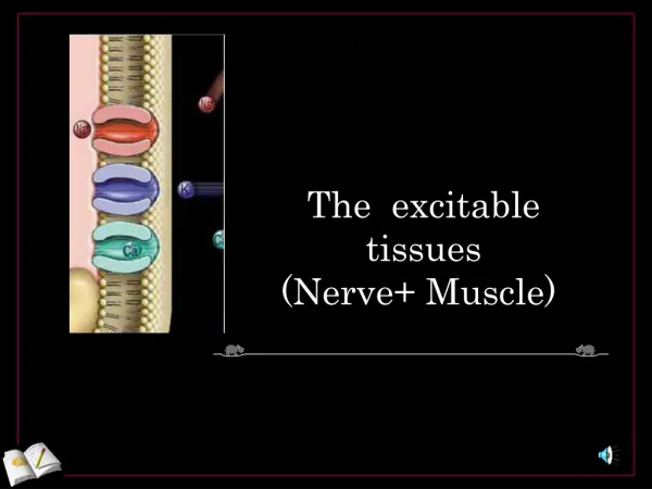
The excitable tissues (Nerve+ Muscle). The nerve. Neuron:- DIF: unit of function of the central nervous system Parts of motor neuron & function of each part: 1- Soma (cell body) 2-Dendrites carry nerve impulses from surroundings to the soma

The excitable tissues (Nerve+ Muscle)
E N D
Presentation Transcript
Neuron:- DIF: unit of function of the central nervous system Parts of motor neuron & function of each part: 1- Soma (cell body) 2-Dendrites carrynerve impulses from surroundings to the soma 3 Axon hillockat which nerve impulses begin 4-Axon & axon terminal
-Histological classification of axons:- 1- myelinated : have myelin sheath (diameter more than 1um) 2- unmyelinated (diameter less than1um ) -type C:postganglionic autonomic &pain fibers
-Myelin sheath is formed by schwann cell which deposit sphingomyelin Functions of myelin sheath 1-insulator 3- increase conduction velocity
RESTING MEMBRANE POTENTIAL DIF: it is potential difference across membrane during rest (without stimulation) Value:- -70 to-90 mv in large nerve fibers ( -ve inside) - -The membrane is polarized
CAUSES: 1- Contribution of K & Na diffusion potential through Na & K leak channels of nerve membrane, CM is more permeable to K than to Na, thus K tends to leak to the out side (down its concentration gradient) carrying positive charge with it. This make the cell interior more negative 2-Active transport of Na & K ions( Na/K pump) 3- Negative ions inside membrane as phosphate & proteins
Causes of RMP: 1. RMP is 100 times more permeable to K+ than Na+. K+ tends to leak out of the cell down its conc gradient, carrying +ve charge with it. (through K leak channels). 2. non-diffusible anions (proteins, sulphate and phosphate ions) cannot leave the cell. 3. very small amount of Na+ diffuses into the cell down its conc gradient. The mb only slightly permeable to Na+. (through Na+ leak channels). 4. Na+-K+ pump maintain conc gradients of K+, and Na+ between the two sides of the mb.
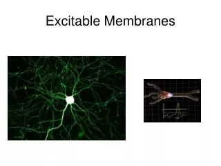
What does it mean when a neuron “fires”? • Firing = excitability = action potential = nerve impulse • Recall resting potential of all cells • High K+ in; high Na+ out • Cell is polarized • Cell overall neg. charge inside due to molecules like proteins, RNA, DNA • Charge measured in millivolts • Potential = difference in charge across PM • Current = flow of charge (ions) from one point to another
Changes that occure through the nerve after stimulation by threshold (effective) stimulus:- 1- Electrical changes (nerve action potential) 2- Excitability changes 3-Thermal changes 4-Chemical changes
The action potential • It is sudden reversal of membrane polarity produced by a stimulus to produce a physiological effect such as: • Transmission of impulse along nerve fibres • Release of neurotransmitters • Muscle contraction • Activation or inhibition of glandular secretion
1- Electrical changes The nerve action potential -It is potential difference along nerve membrane after stimulation by threshold (effective)stimulus -oscilloscope to measure rapid changes in membrane potential -Nerve signals (impulses) are transmitted as nerve action potentials conducted along the nerve fiber as a wave of depolarization to its end -The factors necessary for nerve action potential arevoltage gated Na & k channels -
Summary of events that causes AP:- 1-Initiation of Action Potential (AP) - -70 to-90 mv is the resting potential - Threshold stimulus open voltage gated Na channels & Na influx rises resting potential from -90 towards zero (gradual depolarization) -as membrane potential raises ---------- open more Na channels & more Na influx (+ve feedback ) until all voltage gated Na channels open.
2-Depolarization occurs & membrane potential reach zero value to reach + 35 mv, -at + 35 mv all Na channels begin to close suddenly( Depolarization ends)
c-Repolarization:- due to high K conductance( flow) to outside (K outflux) by openning of all voltage gated K channels (causes negativity inside -
Hyperpolarization: Why? • Na-K pump now start to move Na out & K in against their concentration gradient, so the RMP is resumed and the membrane is ready for another stimulus
The action potential (cont.)*** Threshold stimulus: If a stimulus is strong enough to move RMP from its resting value (-70mV) to the level of (-55mV) which leads to production of an AP
Subthreshold stimulus: Stimulus that result only in local depolarisation
All or nothing principle:- - Once threshold value for excitation is reached a full AP produced ,its intensity can not increased by increasing stimulus intensity ( suprathreshold)
What happens after an action potential? • Refractory period: few millisecs • Time during which can’t stimulate neuron a second time • Happens until recovery of resting potential • Two stages • Absolute refractory period • No new action potential possible • Relative refractory period • Can trigger new action potential if stimulus is very strong
Propagation of action potential 1- in myelinated nerve fibers:- Saltatory conduction ( jumping) Value:- 1-↑ velocity of conduction of nerve impulses 2-Conserve energy for axon because only nodes depolarize
How do action potentials travel down the axon? • Myelinated sheaths • Many times faster transmission • Action potential skips from one node of Ranvier to the next • Called saltatory conduction • http://www.blackwellpublishing.com/matthews/actionp.html
2- Non- myelinated nerves:- (local circuits)=point to point -depolarization pass by local circuits. -
What else influences speed of action potential? .Axon diameter -The larger the diameter, the faster the speed of transmission -Less resistance to current flow with larger diameter Slower transduction Faster transduction
What happens if myelination is lost? • Multiple sclerosis • Autoimmune disease • Usually young adults • Blindness, problems controlling muscles • Ultimately paralysis • Immune system attacks myelin sheaths and nerve fibers • Scar tissue (scleroses) replaces some damaged cells • Other now unmyelinated axons sprout Na+ channels • Accounts for sporadic nature of disease?
Synaptic transmission * • Synapse is the junction between two neurones where electrical activity of one neurone is transmitted to the other
Steps involved • AP at the synaptic knob -----» Ca channels open (increase Ca permeability) -----» • release of neurotransmitter (NT) from synaptic knob to synaptic cleft -----» • NT combines with specific receptors on the other membrane -----» postsynaptic potential -----» AP will result
Chemical Signals One neuron will transmit info to another neuron or to a muscle or gland cell by releasing chemicals called neurotransmitters. The site of this chemical interplay is known as the synapse. An axon terminal (synaptic knob) will abut another cell, a neuron, muscle fiber, or gland cell. This is the site of transduction – the conversion of an electrical signal into a chemical signal.
Synaptic Transmission An AP reaches the axon terminal of the presynaptic cell and causes V-gated Ca2+ channels to open. Ca2+ rushes in, binds to regulatory proteins & initiates NT exocytosis. NTs diffuse across the synaptic cleft and then bind to receptors on the postsynaptic membrane and initiate some sort of response on the postsynaptic cell.
NEUROMUSCULAR JUNCTION AND NEUROMUSCULAR TRANSMISSION OF NERVE ACTION POTENTIAL
-synaptic gutter has subneural folds to increase surface area . Has Ach gated channels (where Ach bind ) at motor end plate in • post-synaptic membrane - synaptic cleft ( filled with ECF & Ach estrase enzyme)
Secretion of acetylcholine(Ach) by nerve terminals ( Ca dependent exocytosis) • 1- AP reach nerve terminal-----open Ca channels------Ca influx------- Ca attract vesicles to nerve terminal membrane , they rupture& release Ach to synaptic cleft( Ca dependent exocytosis)
2- Ach bind to channels--------- then it open 3-, Na flow to inside4-Destruction of Ach by Ach estrase enzymeinto choline & acetate go to nerve terminal to be re-used
Drugs that act on the neuromuscular junction 1-Drugs that act on muscle fiber by Ach like action:- METHACHOLINE- CARBACOL- NICOTINE they act for minutes or hours—as they do not destructed by Ach estrase enzyme . 2-Drugs that block transmission at neuromuscular junction:- CURARE & CURARIFORM likedrugs. act by competitive inhibition to Ach at its receptors & can not cause Depolarization.
3-Drugs that stimulate transmission at neuromuscular junction by inactivation of Ach estrase enzyme:- • A-Neostigmine ,prostigmine andphysostigmine:- inactivates Ach estrase enzyme temporarly • b- di-isopropyl –florophosphate( nerve gas poison) inactivates Ach estrase enzyme for days & weeks -------death because of respiratory muscle spasm