Download
1 / 71
770 likes | 1.52k Vues
Orthopedic Pathology. Arthritis. Osteoarthritis Infectious Rheumatoid. Dislocations. Subluxation partial Luxation complete. Bucket handle tear. Joint mice. Knee Joint. Osteomalacia Osteomyelitis. Osteoporosis Primary Secondary. Osteopathies. Halux valgus Halux varus.

E N D
Arthritis • Osteoarthritis • Infectious • Rheumatoid
Dislocations • Subluxation • partial • Luxation • complete
Bucket handle tear Joint mice Knee Joint
Osteomalacia Osteomyelitis Osteoporosis Primary Secondary Osteopathies
Haluxvalgus Haluxvarus Hammer toe Bunion Toe Deformities
Coxavalga Coxavara Genuvalgum Genuvarum Hip Deformities
Talipes valgus Talipes varus Congenital Anomalies
Compound (open-pierced through the skin) Simple (closed) Direction Linear Spiral Oblique Transverse Intra-articular Comminuted Depressed Fracture line Complete Incomplete Green stick Miscellaneous Colles’ Potts Stellate Impacted Avulsion Spontaneous (pathologic) Fractures
Malignant Ewing’s Tumor Multiple Myeloma Osteosarcoma Benign Chondroma Giant cell tumor Osteoma Neoplasms
8-12 weeks Complete immobilization (internal or external fixation) In proper alignment 5 stages Inflammation 2 days Hematoma-foundation for proliferation Cellular proliferation Macrophages debride Fibrin mesh seals edges of site-fibroblastic and capillary in growth Callus formation 3-4 weeks On outer surface of bone by collagen producing osteoblasts and fibroblasts Bone fragments grow to bridge gap Ossification 2 weeks -4 months Matrix of osteoblasts calcifies and begins to accept mineral deposits Remodeling 1 year Continuous resorption and reforming of bone Normal Bone Healing
Pathological Bone Healing • Causes • Poor immobilization of fracture • Disrupts the hematoma • Distraction of bone fragments • Bone contact does not occur • Gap fills with granulation tissue delaying healing • Interposition of soft tissue • Granulation tissue in gap of distracted bone fragments • Soft tissue grows over both ends of the bone • Deficient blood supply • A vascular necrosis-capillary network cannot be reestablished • Infection • Compound fractures break skin allowing microbes to enter bone and soft tissue • Osteomyelitis-serious bone infection
Pathological Bone Healing-cont. • Delayed union-increased healing time for fractures • Non-union-fractured bone ends don’t unite • Mal-union-fracture heals in improper position and alters mechanical function of the bone • Compartment syndrome-increase in pressure within a closed space • Permanent nerve damage from compression within a fascial compartment. Nerves and vessels are compressed • Necrosis begins in 2-4 hours; irreversible in 12 hours • Usually the forearm or tibia • Fractured bones, casts that are too tight, or intracompartmental bleeding • Damage of other soft tissue by bone fractures • Tendons and ligaments in avulsions (small piece of bone) • Visceral damage from pelvic or rib fractures
Casts Common method for immobilization of a fracture Closed reduction or post operative Materials Fiberglass-inexpensive and light weight Plaster-heavy Supplies for application Bucket with warm water Webril and stockinette Heavy scissors Hardens quickly once wet-maintain position until hard Sticky-gloves Splints Allow for swelling Bivalve cast possibility Materials Orthoglass Sheets of fiberglass Sheets of plaster Supplies for application Bucket with warm water Webril and stockinette Heavy scissors Ace bandage Hardens quickly once wet-maintain position until hard Replaced by a cast in 48 hrs after swelling subsides Casts and Splints
Types of Casts • Short arm-wrist • Long arm-elbow • Short leg-ankle • Long leg-knee • Hip spika-hip or pelvis • Body jacket-spine
Diagnostics • Range of motion-Degree joint can move in any direction • Lachman and Drawer-Ligament stability of knee • Sulcus test-ligament stability of shoulder • Analysis of synovial fluid-osteoarthritis, rheumatoid arthritis (RA), and gout differentiation • Antinuclear antibody-detects autoimmune disease such as lupus or RA • Bence Jones Protein-malignant plasma cells in multiple myeloma. Urine test. • Cultures-infections such as osteomyelitis and septic arthritis • Erythrocyte sedimentation rate (ESR)-rate at which RBC fall to bottom of test tube-fall faster during inflammation. Diagnosis of RA and ankylosing spondylitis. • Human Leukocyte Antigen B27 (HLA)-protein found on WBC-ankylosing spondylitis • Latex fixation test (agglutination tests)-RA detects RA factors • Rheumatoid factors (RF)-RA measure RF in blood • Serum alkaline phosphatase (SAP)-necessary to build new bone. Increased amounts mean bone disease-Multiple myeloma, osteomalacia, osteogenic sarcoma, RA • Serum and urinary calcium and phosphorus-osteoporosis or osteomalacia • Serum urate-gout • Urinary uric acid-gout
Radiology and Diagnostic Imaging • Arthrography-X-ray of joint using contrast medium • Bone Scans-Image of bone after IV injection of Tecnetium (radioactive dye)-picked up by bone with abnormal metabolic activity, detects tumors • Computed Tomography (CT)-X-ray of varying depths. Multiple x-rays at multiple angles. Cross sections (slices) of structures. • Magnetic Resonance Imaging (MRI)-Strong magnetic field produces images best for soft tissues • Skeletal X-rays-simple x-ray of bone
Clinical Procedures • Arthrocentesis-aspiration of synovial fluid-gout, RA, hematoma, infection • Arthroscopy-Visual inspection of joint cavities using a scope. Repair of many conditions is possible using the scope. • Aspiration of Bone Marrow-Withdrawal of bone marrow for diagnosis of blood abnormalities or multiple myeloma • Biopsy-Removal of a piece of bone for pathologist examination-tumors, chronic infection-osteomyelitis
Skull-22 bones Cranium-8 Protects the brain Sinuses reduce weight of skull Facial bones-14 Mandible is only moveable bone of skull Vertebral column Supports head and trunk and protects spinal cord Types Cervical-7 Thoracic-12 Lumbar-5 Sacral-5 Coccyx-1 Bones of the Thoracic Region Ribs Protect Lungs and heart First 7 are true ribs and join the sternum Next 5 are false 3 join cartilage of 7th rib 2 float Sternum-3 sections Manubrium, middle body, xiphoid process Clavicles-braces scapula-AC joint Scapula-shoulder blade Glenoid fossa articulates with head of o Axial Skeleton
Shoulder • AC joint • Rotator cuff- 4 muscles provide strength and stability to shoulder • Infraspinatus, teres minor, subscapularis, and supraspinatus • The 4 tendons insert onto the capsule of the humeral head allowing movement
Arms • Arms • Humerous • Superior • Head, neck, tuberosities • Articulates with Glenoid fossa • Distal • Lateral condyle (capitulum)-articulates with the radius • Medial condyle (troclea)-articulates with ulna • Radius and Ulna • Radius rotates around ulna • Proximal portion of ulna (troclea) articulates with the humerous
Hand and Wrist • Carpals-wrist bones • Distal row-radius to ulna • Trapezium, trapezoid, capitate, hamate • Proximal row-radius to ulna • Scaphoid (navicular), lunate, triquetrum, pisiform • Metacarpals-palm bones • Phalanges-digits-fingers and thumb • #1 is thumb-#5 is pinky
Hip and Pelvis • Pelvis • Sacrum, coccyx, and pelvic girdle • Fusion of 3 bones-ilium, ischium, and pubis • Known as the ox coxae or innominate bone • Iliac crest is primary site of bone grafts • Hip • Acetabulum-cup of ileum for ball and socket joint • Femoral head is the ball • Iliofemoral ligament connects the acetabulum with femoral head
Knee • Knee • Distal femur-lateral and medial condyles • Patellar grove for articulation with patella • Sesamoid bone of the quadriceps tendon • Tibial condyle • Meniscus cushions the articulation of femur and tibia • Thick crescent shaped pads of cartilage • Tendons-gracillus, patellar • Ligaments • Anterior cruciate ligament (ACL) • Posterior cruciate ligament (PCL) • Lateral and medial collateral ligaments (LCL) and (MCL)
Ankle • Tibia-larger and stronger bone of the shin • Sits medial to the fibula • Tibia articulates distally with the talus bone • Medial prominence of the tibia is the medial malleolus • Fibula is non-weight bearing • Lateral malleolus • Talus bone is distal articulation of ankle • Tri-mallelar ankle fracture involves medial and lateral malleoli and talus
Foot • 7 tarsal bones • Calcaneus, navicular, cuboid, medial cuneiform, middle cuneiform, and lateral cuneiform • 5 metatarsals • Phalanges • 3 per toe except big toe (#1)
Positioning Devices • Lower Extremity • Foot prep bar • Sand bags • Fracture table • Upper Extremity • Beach Chair • Traction Bar • Beanbag • Finger traps • Back • Wilson frame • Andrews frame
Other Equipment • Pneumatic Tourniquets • Exsanguination • Cuff • Double or single • Time • 1 hr upper extremity • 1 ½ hr lower extremity • Traction • Lasers • Radiography • Portable • C-arm • Air flow • Laminar flow • Space suits
Other equipment • Continuous Passive Range-of Motion Machines (CPM) • Decreases pain and swelling • Reduces joint stiffness • Inhibits adhesions • Early mobility • Decreases muscle wasting • Transcutaneous Electric Nerve Stimulation (TENS unit) • Stimulates large sensory nerve fibers to suppress pain • Electrical Stimulation of Bone-stimulates osteogenesis and can inhibit bacterial growth • Saws, Drills, Reamers-electric, gas or battery power • Safety switch, weight, • Oscillating or reciprocating • Arthroscopic Equipment-joints • Monitor, light source, arthroscopy pump and shaver, camera, VCR and photo system
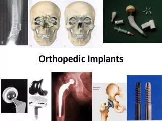
Implants • Screws, plates, wires, pins, IM rods, Joint components • Tracking • Number, type, size, and serial numbers • Alloys-must match metals or corrode • Titanium, stainless steel, cobalt-chromium • Once they go in the patient they are thrown out. • Joints are never re-sterilized or dropped
Other Supplies • Methyl Methacrylate-bone cement • Stabilizes joint components • Tech mixes powder and liquid • Exhaust fumes • Irritate mucous membranes and may be toxic to the liver • Pharmacology • Antibiotics-IV or irrigation • Hemostatic agents-gelfoam, avitene, thrombin, bone wax • Steroids
Done in combination with other procedures Fill a cavity or promote fusion Iliac crest most common-cancellous and cortical Separate drape and instrument set-up Gouges, osteotomes, curettes, mallet Procedure Incision-10 blade Dissection Curved Mayo scissors, rat tooth pick-up Retractors Dissect fascia and periosteum Large elevator (key or cobb) Procure graft Osteotome and mallet Curettes, gouge, rongeur Keep graft in saline or blood Possible bone wax for hemostasis Close Bone Grafting
Equipment Video, tourniquet, fluid pump, shaver, post or other fixation device, tourniquet Instruments Arthroscopy set, scope, graspers and baskets May drop foot of table Lots of cords and tubing, prime fluid tubing May use a spinal needle for incision placement Lateral release, chondral debridement, plica band excision Procedure Incision #11 blade Irrigation cannula is placed and trocar is removed Attach irrigation tubing and insert scope 2nd incision #11 blade and blunt trocar Explore joint Nerve hook Repair Shaver and baskets Close Knee Arthroscopy
Bucket handle tear Try to preserve as much of the cartilage as possible Meniscectomy or repair Follow procedure for arthroscopy up to repair May require post operative physical therapy Procedure Perform Diagnostic arthroscopy Free torn piece of meniscus Nerve Hook Grasp torn segment Arthroscopic grasper Place meniscal cannulas and advance long needle suture Incision into poplietal space #15 blade, metz, army-navy Advance needles and tie suture Close Arthroscopic Meniscal Tear Repair
Attaches medial edge of lateral femoral condyle in notch and interspinous area of the tibia Stabilizes the knee Injured in basketball, football or skiing Valgus twisting injury Allograft or autograft Patellar tendon, gracillus, iliotibial band, or semitendinosus Instruments Ortho set, arthroscopy set, scope equipment, ACL system, saw and drill set up, bone fixation devices Screws, staples, washers Extremity drape Procedure Arthroscopic exam and debridement Scope, shaver, baskets Notchplasty-widens inter condylar notch Arthroscopic bur, osteotome, rasp Graft harvest Incision/dissection anterior tibia #15 blade, senns, elevator, metz, rat tooth Tendon stripper (soft tissue only), #2 or 5 ethibond Saw (bone-tendon-bone), rongeur, small drill bit, #2 0r 5 ethibond Graft prep jig Measure diameter of graft Arthroscopic Anterior Cruciate Ligament Repair
Tibial Tunnel Tibial aiming device is placed at notch through Arthroscopic port Guide pin is advanced through anterior tibia through tibial spine Drill and pin Larger of 2 drill bit dimensions for graft is advanced over guide pin Drill and drill guide Femoral Tunnel Long guide pin with eye is advance through intercondylar notch through skin of thigh Army-navy and pliers Smaller diameter drill bit is advance over guide pin through tibial tunnel. It does not penetrate femur on distal side Drill and guide Leave guide pin in place remove drill Graft Insertion and fixation Thread ethibond on smaller diameter end of graft through eye of guide pin. Pull guide pin through skin of thigh causing graft to advance to position inside femoral tunnel Pliers Insert screw guide pin through arthroscopic port Advance no-profile screw into notch over graft Screw driver Keep tension on distal ethibond Fix distal portion of graft to anterior tibia Staples or screw Trim graft at tibia Close ACL continued