
Uploaded by
audrey-holman
1 SLIDES
121 VUES
10LIKES
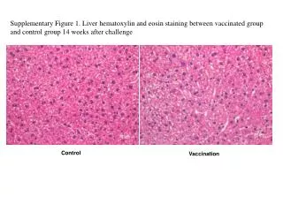
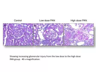
Liver Histological Analysis: Comparing Vaccinated and Control Groups Post-Challenge
DESCRIPTION
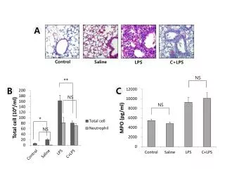
This supplementary figure illustrates the liver histopathology using hematoxylin and eosin staining in both vaccinated and control groups, evaluated 14 weeks after the challenge. It provides critical insights into the hepatic response to vaccination, highlighting any morphological differences or damage within liver tissues. The figure serves to compare the immune response in the vaccinated group against the control, offering evidence of the vaccine's protective or reactive effects on liver health post-exposure.
Download
1 / 1
Télécharger la présentation 
Liver Histological Analysis: Comparing Vaccinated and Control Groups Post-Challenge
An Image/Link below is provided (as is) to download presentation
Download Policy: Content on the Website is provided to you AS IS for your information and personal use and may not be sold / licensed / shared on other websites without getting consent from its author.
Content is provided to you AS IS for your information and personal use only.
Download presentation by click this link.
While downloading, if for some reason you are not able to download a presentation, the publisher may have deleted the file from their server.
During download, if you can't get a presentation, the file might be deleted by the publisher.
E N D
Presentation Transcript
Supplementary Figure 1. Liver hematoxylin and eosin staining between vaccinated group and control group 14 weeks after challenge Control Vaccination


More Related