Download
1 / 19
230 likes | 2.04k Vues
Sodium dodecyl sulfate-Polyacrylamide gel electrophoresis (SDS-PAGE). Irene Goh Rosarine Metusela. Objectives. To use the SDS PAGE analytical procedure to identify and/or isolate the following proteins: •Ovalbumin •Casein •Gluten

E N D
Sodium dodecyl sulfate-Polyacrylamide gel electrophoresis (SDS-PAGE) Irene Goh Rosarine Metusela
Objectives • To use the SDS PAGE analytical procedure to identify and/or isolate the following proteins: •Ovalbumin •Casein •Gluten • To be able to understand the principles of gel electrophoresis • To apply and follow safety procedures while carrying out the experiment
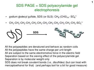
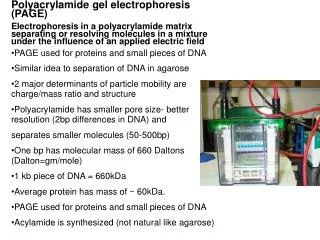
What is SDS-PAGE? • Based on the migration of charged molecules in an electric field • Separation technique • Uses the Polyacrylamide gel as a “support matrix”. The matrix inhibits convective mixing caused by heating and provides a record of the electrophoretic run. • Polyacrylamide is a porous gel which acts as a sieve and separates the molecules
Role of SDS • Denatures proteins by wrapping around the polypeptide backbone. • SDS binds to most proteins in amount roughly proportional to molecular weight of the protein-about one molecule of SDS for every two amino acids (1.4 g SDS per gram of protein) (Lehninger Principles of Biochemistry). • In doing so, SDS creates a large negative charge to the polypeptide in proportion to its length
Role of SDS (cont…) • SDS also disrupts any hydrogen bonds, blocks many hydrophobic interactions and partially unfolds the protein molecules minimizing differences based on the secondary or tertiary structure • Therefore, migration is determined not by the electrical charge of the polypeptide, but by molecular weight. • The rate at which they move is inversely proportional to the molecular mass • This movement is then used to determined the molecular weight of the protein present in the sample.
Procedure: materials • 1.A Mighty Small II, SE 260 Mini-Vertical Gel Electrophoresis Unit • 2.0.5 TrisCl, pH 6.8 solution • 3.10% SDS solution • 4.Sample treatment buffer • 5.SDS glycine running buffer • 6.β-Mercaptoethanol solution • 7.Brilliant Blue R concentrate • 8.Destaining solution • 9.Precast polyacrylamide separating gel • 10.Fine tipped microsyringe • 11.Protein samples (ovalbumin, casein, and gluten)
Procedure: solutions • 0.5M TrisCl, pH 6.8 (4X Resolving gel buffer) • 10% SDS solution • 2X Sample treatment buffer • SDS glycine running buffer • Destaining solution
Procedure: electrophoresis unit • Initial preparation-wash the unit • Preparing the gel sandwich(es): • ensure that the plates are completely polymerized before loading • Install the gel sandwhich(es) into the unit before loading any of the protein samples. • Loading the protein samples: • Dry sample: add equal volumes of treatment buffer solution, and deionised water to achieve the required concentration. Heat in a tube, in boiling water for 90 seconds
Procedure: electrophoresis unit • Fill upper buffer chamber with running buffer • Using a fine-tipped microsyringe, load the treated protein samples into the wells so that the volume in each well is raised by 1mm • Fill the lower buffer chamber
Procedure: running the gel • Place the safety lid on before plugging in the leads of the unit to the power supply. • Run the gel at 20mA per gel, using a constant current • When it reaches the bottom of the gel, the run is complete • Turn off the power supply, and disconnect the leads, before removing the safety lid
Procedure: running the gel • Carefully remove the gel(s) from the plates • Lay it into a tray of staining solution for about 10 minutes. • Remove the gel carefully and place it in between two layers of transparencies, cut along the edges of the gel and analyse the results.
Results and discussion • The results discussed here is, the sample results which was provided by the supervisor
Results and discussion • the relationship between the logarithm of the standards and the relative distance travelled by each protein through the gel is linear • The equation of the line was obtained and used to calculate the relative molecular weights (Mr) of the samples in lanes b-l of the gel • x = (y + 1.7679)/0.4785 x – Mr y – Relative distance travelled by the sample in centimetres
Results and discussion • From the molecular weights obtained for the proteins to be analysed in the experiment: • Cassein = 24,000 Da • Ovalbumin = 46,000 Da • Gluten = 20,000 – 11,000,000 Da • It would be expected that the relative molecular weights of these proteins, would be close their respective theoretical values shown above.
Conclusion • SDS PAGE is a useful method for separating and characterising proteins, where a researcher can quickly check the purity of a particular protein or work out the different number of proteins in a mixture. • Since we did not obtain results for the experiment, • we have to rely on sample results • Cannot validate the experimental technique