Thorax Lungs
Thoracic Cage. Bony structure in a conical shape, narrower at the top

Thorax Lungs
E N D
Presentation Transcript
1. Thorax & Lungs
2. Thoracic Cage Bony structure in a conical shape, narrower at the top & wider at the bottom
Sternum
12 pairs of ribs � 1-7 attach to sternum; 8,9,10 attach to costal cartilage; 11 & 12 floating
12 thoracic vertebrae
Diaphragm � musculotendinous septum that separates the thorax from the abdomen
3. Anterior Landmarks Suprasternal notch
Sternum � manibrium, body, xiphoid process
Manubriosternal angle � or Angle of Louis. Continues with second rib. Marks where the trachea bifurcates into right & left bronchi; marks the upper border of the heart
Costal angle � right & left costal margins meet here. Usually less than 90 degrees
4. Posterior Landmarks Counting ribs & intercostal spaces more difficult on posterior b/c muscle & soft tissue
Vertebra prominens � C7, then T1
Spinous processes � count down the knobs on the vertebrae
Scapulae end at 7th or 8th rib
12th rib can feel its free tip
5. Reference Lines
Anterior chest � midsternal line, midclavicular line
8. Mediastinum � middle section of thoracic cavity containing esophagus, trachea, heart, & great vessels
Pleural cavity � on either side of the mediastinum, contains lungs
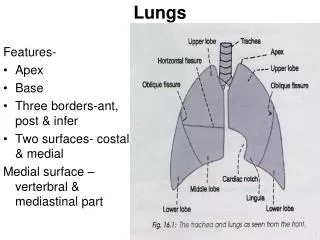
9. Lung borders Anterior � apex is highest point, 3-4cm above inner 1/3 of clavicles. Base rests on diaphragm at 6th rib midclavicular line
Posterior � apex is near C7. Base is at T10
10. Lung lobes Right lung has 3 lobes & is shorter than left b/c of liver placement
Left lung has 2 lobes & is narrower than right b/c heart bulges
Lobes are separated by fissures
Posterior chest is almost all lower lobe. Upper lobes extend down to T3
11. Pleurae Thin, slippery serous membrane that forms an envelope between lungs & chest wall
Visceral pleura lines outside the lungs
Parietal pleura lines inside the chest wall
Inside pleural cavity is pleural fluid that lubricates for smooth movement during respiration. A vacuum with negative pressure that holds the lungs tight against chest wall
12. Trachea & Bronchial Tree Trachea � anterior to esophagus, 10-11 cm long. Begins at cricoid cartilage in neck & bifurcates just below sternal angle into R & L bronchi. Posteriorly, bifurcates at T4-T5.
Right bronchus is shorter, wider, & more vertical .
Bronchi lined with goblet cells that secrete mucus that traps particles & lined with cilia that sweeps particles up where they can be swallowed. Made up of elastic, connective tissue to expel air from lungs
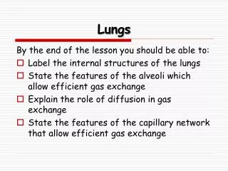
13. Acinus Functional respiratory unit that consists of bronchioles, alveolar ducts, alveolar sacs, & alveoli
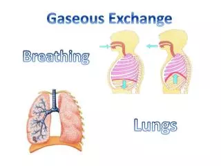
Gaseous exchange occurs across the respiratory membrane in the alveoli
14. Mechanics of Respiration 4 major functions
Supplying oxygen to the body for energy production
Removing carbon dioxide as a waste product of energy reactions
Maintaining homeostasis (acid-base balance) of arterial blood
Maintaining heat exchange (less important in humans)
15. O2/CO2 exchange maintains pH balance of the blood within its normal range
Control of respirations is involuntary, by respiratory center in brain stem (pons & medulla). Normal stimulus to breathe is an increase in carbon dioxide in the blood (hypercapnia)
16. Changing chest size Air comes in � chest size increases
Exhale � chest size decreases
Vertically � diameter will move up or down
AP diameter � increases or decreases with elevation or depression of ribs
17. Inspiration vs. Expiration Inspiration: increasing size of thoracic container creates a slight negative pressure so air rushes in. Diaphragm descends, lengthens vertical diameter, sternum & ribs elevate thus increasing anteroposterior diameter
Expiration is passive: diaphragm relaxes, elastic forces in lungs force the air out
18. Subjective Data Cough
SOB
Chest pain with breathing
Hx of respiratory infections
Hx smoking
Environmental exposure
Self-care behaviors � flu shot, TB skin test