Download
1 / 26
260 likes | 859 Vues
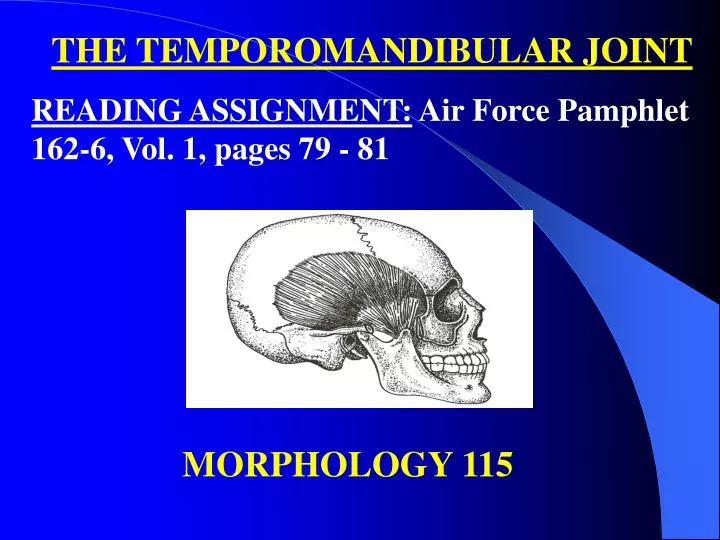
THE TEMPOROMANDIBULAR JOINT READING ASSIGNMENT: Air Force Pamphlet 162-6, Vol. 1, pages 79 - 81. MORPHOLOGY 115.

E N D
THE TEMPOROMANDIBULAR JOINT READING ASSIGNMENT: Air Force Pamphlet 162-6, Vol. 1, pages 79 - 81 MORPHOLOGY 115
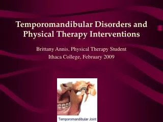
Overview: The right and left temporomandibular joints are the two places where the mandible connects with the rest of the skull. In general terms, the temporomandibular joint is formed by the glenoid fossa and articular eminence of the temporal bone and by the condyle of the mandible. The fossa and eminence are separated from contact with the condyle by an articular disk.
Temporomandibular Joint Structure: Glenoid Fossa: The glenoid fossa is a deep hollow on the under surface of the zygomatic process of the temporal bone. The base of the zygomatic process is the place where the process originates from the central mass of the temporal bone. The condyle stays in the fossa during ordinary opening and closing (hinge) movements.
Articular Eminence: The articular eminence is a ramp-shaped prominence which extends forward and downward from the anterior boundary of the glenoid fossa. During forward (protrusive) movements of the entire mandible, both condyles leave their fossa and move onto eminences. In lateral movements, one condyle usually stays in a fossa and the other condyle moves out of the fossa onto its eminence.
Condyle: The condyle is the oval- or kidney-shaped structure found on the end of the condyloid process.
Articular Disc: The articular disc is a pad of tough, flexible fibrocartilage situated between the condyle and the glenoid fossa. The disc is a shock-absorbing mechanism. When the condyle moves out onto the articular eminence, the disc travels with it. NOTE: Another name for the articular disc is the meniscus.
Synovial Cavities: The synovial cavities are also referred to as the upper and lower joint compartments: 1. Upper. The upper synovial cavity is found between the top of the disc and the glenoid fossa. 2. Lower. The lower synovial cavity is found between the bottom of the disc and the condyle of the mandible.
Synovial Membrane and Associated Synovial Fluid: The synovial membrane is the lining of a synovial cavity. The cells of the lining make a lubricating liquid called synovial fluid.
Capsule: The capsule is the major ligament of the temporomandibular joint. This ligamentous sleeve or capsule originates from the entire rim of the glenoid fossa and articular eminence, attaches to the edges of the articular disc, and passes to insert around the rim of the condyle. The capsule holds the disc in place between the condyle and the fossa, it retains the synovial fluid in the upper and lower joint compartments, and it acts to prevent dislocation of the mandible. Some authors of anatomy texts mention a temporomandibular ligament, which is an anterior thickening of the capsule, not a separate ligament.
Auxillary Ligaments: Auxillary ligaments generally act to restrict the condyle to a normal range-of-movement and prevent dislocation. 1. Stylomandibular Ligament. The stylomandibular ligament originates on the styloid process of the temporal bone and inserts on the posterior border of the ramus near the angle. 2. Sphenomandibualr Ligament. The sphenomandibular ligament originates on the spine of the sphenoid bone and inserts on the anterior-superior of the mandibular foramen (lingula). The mandibular foramen is found on the internal surface of the ramus of the mandible.
Basic Mandibular Movements Opening and Closing: From a position of centric relation, pure hinge movements are possible in opening and closing. In a hinge movement, the condyles rotate within the glenoid fossa. Opening and closing movements, where the measured distance between maxillary and mandibular incisors is greater than 25mm, result in combined rotation and translation of the condyles. Translation occurs whenever a condyle leaves the glenoid fossa.
Protrusion and Retrusion: Protrusion is when the mandible moves forward and both condyles leave their respective fossae and move down their eminences. The opposite process is called retrusion. Protrusion and retrusion are translatory movements.
Right and Left Lateral: 1. Working side: The side toward which the mandible moves. When the mandible moves laterally, the condyle on the working side stays in its fossa, rotates and moves laterally. 2. Balancing side: The side opposite the working side. In a lateral movement, the balancing side condyle leaves the fossa and moves forward and down the eminence, and medially.