Download
1 / 1
10 likes | 167 Vues
Growth of Self-Assembled BaTiO 3 Nanodots using Tensile Strain. Timothy A. Morgan, Zhaoquan Zeng, Robert J. Sleezer, Gregory J. Salamo. Department of Physics, University of Arkansas, Fayetteville, Arkansas

E N D
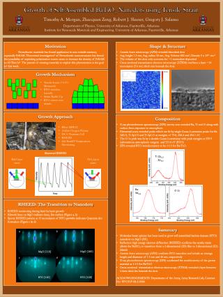
Growth of Self-Assembled BaTiO3Nanodots using Tensile Strain Timothy A. Morgan, Zhaoquan Zeng, Robert J. Sleezer, Gregory J. Salamo Department of Physics, University of Arkansas, Fayetteville, Arkansas Institute for Nanoscale Materials and Engineering, University of Arkansas, Fayetteville, Arkansas Motivation Ferroelectric materials has found application in non-volatile memory, especially FeRAM. Theoretical investigation1of Ferroelectric nanostructures has found the possibility of exploiting polarization vortex states to increase the density of FeRAM to 60 Tbit/in2. The pursuit of creating materials to exploit this phenomenon is the goal of this work. • Shape & Structure • Atomic force microscopy (AFM) revealed disorderd dots • Avg. height 1.3 nm, Avg. radius 18 nm, Avg. Volume 882 nm3, Density 5 x 1011 cm-2 • The volume of the dots only accounts for ~1 monoalyer deposited • Cross-sectional transmission electron microscopy (XTEM) confirms a layer ~14 monolayers (5.6 nm) thick exis beneath the dots. Growth Mechanism • Tensile Strain (+5.5% Mismatch) • BTO stretches laterally • Strain Builds Up • BTO relaxes into islands Growth Approach Shuttered RHEED • Composition • X-ray photoelectron spectroscopy (XPS) survey scan revealed Ba, Ti and O along with carbon from exposure to atmosphere. • Elemental scans revealed peaks which are fit by single Gauss-Lorentzian peaks for Ba 3d5/2, Ti 2p3/2 and Ti 2p1/2 at energies of 79.8, 458.4 and 454.1 eV. • The O 1s peak was fit by a double Gauss-Lorentizian with peak energies at 529.9 (adventitious atmospheric oxygen) and 531.8 eV (BTO) • XPS revealed BTO stoichiometery to be 1:1:3 for Ba:Ti:O. • Riber MBE32 • Addon Oxygen Plasma • DCA Titanium Cell • RHEED • kSa BandiT Tempearture Monitoring BaO layer starts TiO2 layer starts b a Ti 2p1/2 2p3/2 MgO [100] MgO [110] c d • RHEED: The Transition to Nanodots • RHEED monitoring during layer by layer growth • Kikuchi lines on MgO indicate clean, flat surface (Figurs a, b) • Spotty RHEED pattern at 15 monolayers of BTO growths indicates Quantum dot formation (Figure c & d) BTO [110] BTO [100] Ba 3d5/2 O 1s • Summary • Molecular beam epitaxy has been used to grow self-assembled barium titanate (BTO) nanodots on MgO (001) • Reflection high energy electron diffraction (RHEED) confirms the tensile strain allows the BaTiO3 to transition from a 2-dimenisonal (2D) film to 3-dimensional (3D) islands • Atomic force microscopy (AFM) confirms BTO nanodots and reveals an average height and diameter of 1.3 nm and 36 nm, respectively • X-ray photoelectron spectroscopy (XPS) confirmed the stoichiometry of the grown material as 1:1:3 for Ba:Ti:O • Cross-sectional transmission electron microscopy (XTEM) revealed a layer between 4-6nm thick lies beneath the dots • ACKNOWLEDGEMENTS: Department of the Army, Army Research Lab, Contract No: W911NF-08-2-0006