Neural Condition: Synaptic Transmission & Membrane Potential
320 likes | 360 Vues
Explore synaptic transmission and resting membrane potential in neurons, covering ion channels, polarization, depolarization, neural integration, and action potentials.

Neural Condition: Synaptic Transmission & Membrane Potential
E N D
Presentation Transcript
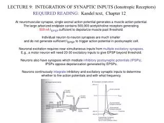
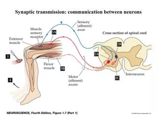
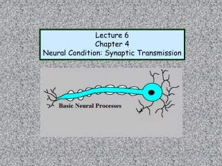
Lecture 6 Chapter 4 Neural Condition: Synaptic Transmission
Chemical Transmission • Transmitting Step: presynaptic cell • release NT • 2. Receptive Step: NT binds postsynaptically • to chemically gated (ion) channel presynaptic postsynaptic
Resting membrane potential: AT REST The differences in electrical charges between the inside of the cell and the outside cell “Polarized”
Why Negative You ASK? IONS Charged Molecules Cl- K+ Na+ A- Na+: Cation (+) K+: Cation (+) Cl-: Anion (-) A-: Anion (-) Ratio of negative to positive is greater inside than outside
Why Unequal Distribution? 4 Factors Equalizers Unequalizers 1. Concentration Gradient (passive) • Random movement of ions • Ions travel “down” their [ ] gradient • higher concentration moves to region of lower concentration • Stops when ions are =
2. Electrostatic Pressure (Passive) Charges • Opposites attract • Like charges repel Hey baby.. What’s your sign? Hey baby.. What’s your sign? Talk to the Membrane.. Na+ Cl- K+ Na+
3. Selectivity of Membrane (Passive) Cell membrane is permeable to certain Ions – controls in & out
4. Sodium Potassium Pump (Energy-Active) For every 2 K+ in 3 Na+ out (transport system)
End Result… Polarization
IONIC BASIS OF THE RESTING MEMBRANE POTENTIAL • General overview • Resting membrane potential is a characteristic feature of ALL cells in the body • Membranes of neurons contain channels that allow the movement of ions into or out of the cell • The permeability of the membrane is determined by the state of these channels – i.e. whether they are open or closed
Neurons have the ability to “gate” their ion channels • (permeability of the membrane to selected ions) • 3 basic types of ion channels: • (i) Passive – each passive channel is identified by the specific ion it allows through it(e.g. K+, Na+ channel) resting membrane potential (ii) Voltage gated – open or close based on the membrane potential action potentials (AP)(iii) Chemically gated – transmitters binding on sites (receptors) on the channels. These channels are important in synaptic transmission
Depolarize the Cell Make more positive How do we get the cell to depolarize?
How do we get the cell to depolarize? EPSP: excitatory post synaptic potential Increases the likelihood that postsynaptic cell will fire • NT binds to chemical gated channel • Ion Channel opens (NA+) • Flood of Na rushes in…why? • Cell becomes depolarize (–70mV) • If reaches “threshold” (-65 mV) • Action Potential (AP)! Will be triggered!!!!
NT binds to chemical gated channel • Ion Channel opens (NA+) • Flood of Na rushes in…why? • Cell becomes depolarize (–70mV) • If reaches “threshold” (-65 mV) • Action Potential (AP)! Will be triggered!!!!
How do we reach threshold? Neural Integration: Cell “adds” up signals over space & time GRADED RESPONSE Spatial Summation: Many synaptic inputs adds up to threshold Temporal Summation: One Input that fires quickly in time serving to build on each other To reach threshold AP triggered
Integration of Signals Figure 8-25: Locations of synapses on a postsynaptic neuron
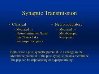
Postsynaptic and Action Potentials Excitatory Postsynaptic Potentials (EPSP) Graded depolarizations Inhibitory Postsynaptic Potentials (IPSP) Graded hyperpolarizations Relationship between EPSP’s, IPSP’s and AP???? All postsynaptic potentials are added together and if enough EPSP’s occur to cause cell to cross threshold, an action potential occurs
Hits Threshold All-or-Nothing! Action Potential: firing of a neuron massive momentary change in the membrane potential from –70mV to ~ +50mV
Figure 3.3 Recordings from a postsynaptic neuron during synaptic activation
Getting back resting membrane potential
Properties of the action potential • AP is triggered by depolarization • Depolar. must exceed threshold value to trigger AP • AP is all-or-none • AP propagates without decrement • AP involves reversal ("overshoot") of membrane potential • AP is followed by refractory period
Voltage Gated Channels in axon open serving to “Propogate” the AP down the axon
AP generated at every single spot all the way down the axon • Voltage gated Channels only at Nodes of Ranvier • AP only generated at “Nodes of Ranvier” less depolarizing “Saltatory Conduction”