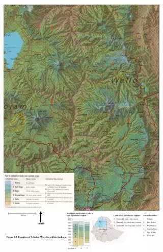
Figure 2.1
“Feet” of leaf beetle (left) and bush fly (right). (From scanning electron micrographs by C.A.M. Reid & A.C. Stewart.). Figure 2.1. The general structure of insect cuticle; the enlargement above shows details of the epicuticle. (After Hepburn 1985; Hadley 1986; Binnington 1993.). Figure 2.2.
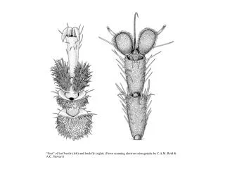

Figure 2.1
E N D
Presentation Transcript
“Feet” of leaf beetle (left) and bush fly (right). (From scanning electron micrographs by C.A.M. Reid & A.C. Stewart.)
Figure 2.1 The general structure of insect cuticle; the enlargement above shows details of the epicuticle. (After Hepburn 1985; Hadley 1986; Binnington 1993.)
Figure 2.2 Structure of part of a chitin chain, showing two linked units of N-acetyl-D-glucosamine. (After Cohen 1991.)
Figure 2.3 The ultrastructure of cuticle (from a transmission electron micrograph). (a) The arrangement of chitin microfibrils in a helicoidal array produces characteristic (though artifactual) parabolic patterns. (b) Diagram of how the rotation of microfibrils produces a lamellar effect owing to microfibrils being either aligned or non-aligned to the plane of sectioning. (After Filshie 1982.)
Figure 2.4 A specialized worker, or replete, of the honeypot ant, Camponotus inflatus (Hymenoptera: Formicidae), which holds honey in its distensible abdomen and acts as a food store for the colony. The arthrodial membrane between tergal plates is depicted to the right in its unfolded and folded conditions. (After Hadley 1986; Devitt 1989.)
Figure 2.5 The cuticular pores and ducts on the venter of an adult female of the citrus mealybug, Planococcus citri (Hemiptera: Pseudococcidae). Enlargements depict the ultrastructure of the wax glands and the various wax secretions (arrowed) associated with three types of cuticular structure: (a) a trilocular pore; (b) a tubular duct; and (c) a multilocular pore. Curled filaments of wax from the trilocular pores form a protective body-covering and prevent contamination with their own sugary excreta, or honeydew; long, hollow, and shorter curled filaments from the tubular ducts and multilocular pores, respectively, form the ovisac. (After Foldi 1983; Cox 1987.)
Figure 2.6 The four basic types of cuticular protuberance: (a) a multicellular spine; (b) a seta, or trichoid sensillum; (c) acanthae; and (d) microtrichia. (After Richards & Richards 1979.)
Figure 2.7 Types of body segmentation. (a) Primary segmentation, as seen in soft-bodied larvae of some insects. (b) Simple secondary segmentation. (c) More derived secondary segmentation. (d) Longitudinal section of dorsum of the thorax of winged insects, in which the acrotergites of the second and third segments have enlarged to become the postnota. (After Snodgrass 1935.)
Figure 2.8 The major body axes and the relationship of parts of the appendages to the body, shown for a sepsid fly. (After McAlpine 1987.)
Figure 2.9 Lateral view of the head of a generalized pterygote insect. (After Snodgrass 1935.)
Figure 2.10 Frontal view of the head and dissected mouthparts of an adult of the European earwig, Forficula auricularia (Dermaptera: Forficulidae). Note that the head is prognathous and thus a gular plate, or gula, occurs in the ventral neck region.
Figure 2.11 Frontal view of the head of a worker honey bee, Apis mellifera (Hymenoptera: Apidae), with transverse section of proboscis showing how the “tongue” (fused labial glossae) is enclosed within the sucking tube formed from the maxillary galae and labial palps. (Inset after Wigglesworth 1964.)
Figure 2.12 Mouthparts of a white butterfly, Pieris sp. (Lepidoptera: Pieridae). (a) Positions of the proboscis showing, from left to right, at rest, with proximal region uncoiling, with distal region uncoiling, and fully extended with tip in two of many possible different positions due to flexing at “knee bend”. (b) Lateral view of proboscis musculature. (c) Transverse section of the proboscis in the proximal region. (After Eastham & Eassa 1955.)
Figure 2.13 Female mosquito mouthparts in (a) frontal view; (b) transverse section. ((a) After Freeman & Bracegirdle 1971; (b) after Jobling 1976.)
Figure 2.14 Mouthparts of adult Diptera. (a) House fly, Musca (Muscidae). (b) Stable fly, Stomoxys (Muscidae). (After Wigglesworth 1964.)
Figure 2.15 Head and mouthparts of a thrips, Thrips australis (Thysanoptera: Thripidae). (a) Dorsal view of head showing mouthparts through prothorax. (b) Transverse section through proboscis. The plane of the transverse section is indicated by the dashed line in (a). (After Matsuda 1965; CSIRO 1970.)
Figure 2.16 Head and mouthparts of a sucking louse, Pediculus (Psocodea: Anoplura: Pediculidae). (a) Longitudinal section of head (nervous system omitted). (b) Transverse section through eversible proboscis. The plane of the transverse section is indicated by the dashed line in (a). (After Snodgrass 1935.)
Figure 2.17 Head and mouthparts of a human flea, Pulex irritans (Siphonaptera: Pulicidae): (a) lateral view of head; (b) transverse section through mouthparts. The plane of the transverse section is indicated by the dashed line in (a). (After Snodgrass 1946; Herms & James 1961.)
Figure 2.18 The mouthparts and feeding currents of a mosquito larva of Anopheles quadrimaculatus (Diptera: Culicidae). (a) The larva floating just below the water surface, with head rotated through 180° relative to its body (which is dorsum-up so that the spiracular plate near the abdominal apex is in direct contact with the air). (b) Viewed from above showing the venter of the head and the feeding current generated by setal brushes on the labrum (direction of water movement and paths taken by surface particles are indicated by arrows and dotted lines, respectively). (c) Lateral view showing the particle-rich water being drawn into the preoral cavity between the mandibles and maxillae and its downward expulsion as the outward current. ((b,c) After Merritt et al. 1992.)
Figure 2.19 Some types of insect antennae: (a) filiform, linear and slender; (b) moniliform, like a string of beads; (c) clavate or capitate, distinctly clubbed; (d) serrate, saw-like; (e) pectinate, comb-like; (f) flabellate, fan-shaped; (g) geniculate, elbowed; (h) plumose, bearing whorls of setae; and (i) aristate, with enlarged third segment bearing a bristle.
Figure 2.20 Diagrammatic lateral view of a wing-bearing thoracic segment, showing the typical sclerites and their subdivisions. (After Snodgrass 1935.)
Figure 2.21 The hind leg of a cockroach, Periplaneta americana (Blattodea: Blattidae), with enlargement of ventral surface of pretarsus and last tarsomere. (After Cornwell 1968; enlargement after Snodgrass 1935.)
Figure 2.22 Nomenclature for the main areas, folds, and margins of a generalized insect wing.
Figure 2.23 A generalized wing of a neopteran insect (any living winged insect other than Ephemeroptera and Odonata), showing the articulation and the Kukalová-Peck nomenclatural scheme of wing venation. Notation as follows: AA, anal anterior; AP, anal posterior; Ax, axillary sclerite; C, costa; CA, costa anterior; CP, costa posterior; CuA, cubitus anterior; CuP, cubitus posterior; hm, humeral vein; JA, jugal anterior; MA, media anterior; m-cu, cross-vein between medial and cubital areas; MP, media posterior; PC, precosta; R, radius; RA, radius anterior; r-m, cross-vein between radial and median areas; RP, radius posterior; ScA, subcosta anterior; ScP, subcosta posterior. Branches of the anterior and posterior sector of each vein are numbered, e.g. CuA1–4. (After CSIRO 1991.)
Figure 2.24 The left wings of a range of insects showing some of the major wing modifications: (a) fore wing of a butterfly of Danaus (Lepidoptera: Nymphalidae); (b) fore wing of a dragonfly of Urothemis (Odonata: Anisoptera: Libellulidae); (c) fore wing or tegmen of a cockroach of Periplaneta (Blattodea: Blattidae); (d) fore wing or elytron of a beetle of Anomala (Coleoptera: Scarabaeidae); (e) fore wing or hemelytron of a mirid bug (Hemiptera: Heteroptera: Miridae) showing three wing areas, the membrane, corium, and clavus; (f) fore wing and haltere of a fly of Bibio (Diptera: Bibionidae). Nomenclatural scheme of venation consistent with that depicted in Fig. 2.23; that of (b) after J.W.H. Trueman, unpublished. ((a–d) After Youdeowei 1977; (f) after McAlpine 1981.)
Figure 2.25 The female abdomen and ovipositor: (a) lateral view of the abdomen of an adult tussock moth (Lepidoptera: Lymantriidae) showing the substitutional ovipositor formed from the extensible terminal segments; (b) lateral view of a generalized orthopteroid ovipositor composed of appendages of segments 8 and 9; (c) transverse section through the ovipositor of a katydid (Orthoptera: Tettigoniidae). T1–T10, terga of first to tenth segments; S2–S8, sterna of second to eighth segments. ((a) After Eidmann 1929; (b) after Snodgrass 1935; (c) after Richards & Davies 1959.)
Figure 2.26 Male external genitalia. (a) Abdominal segment 9 of the bristletail Machilis variabilis (Archaeognatha: Machilidae). (b) Aedeagus of a click beetle (Coleoptera: Elateridae). ((a) After Snodgrass 1957.)