Neoplasia
670 likes | 935 Vues
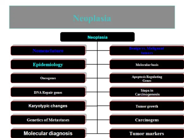
Neoplasia. Nomenclature. Benign: - oma (adenoma, fibroma) Malignant: carcinoma ( epithelial origin ) (adenocarcinoma) sarcoma ( mesenchymal origin ) (fibrosarcoma). Adenoma. Adenoma. Papilloma. Polyp. Non-Neoplastic Proliferation:. Controlled & Reversible Hypertrophy – Size

Neoplasia
E N D
Presentation Transcript
Nomenclature • Benign: -oma (adenoma, fibroma) • Malignant: • carcinoma (epithelial origin) • (adenocarcinoma) • sarcoma (mesenchymal origin) • (fibrosarcoma)
Non-Neoplastic Proliferation: • Controlled & Reversible • Hypertrophy – Size • Hyperplasia – Number • Metaplasia – Change • Dysplasia – Disordered
Neoplastic Proliferation: Uncontrolled & Irreversible • Benign • Localized, non-invasive. • Malignant (Cancer) • Spreading, Invasive.
Dysplasia • Loss of architectural organization and orientation and a loss of cell uniformity in tissue. • Dysplasia is a non-neoplastic proliferation. • Dysplasia may or may not progress to cancer.
Here, there is normal cervical squamous epithelium at the left, but dysplastic squamous epithelium at the right. Dysplasia is a disordered growth of epithelium, but still confined to the epithelium. Dysplasia is still reversible.
At high magnification, the normal cervical squamous epithelium at the left merges into the dysplastic squamous epithelium at the right in which the cells are more disorderly and have darker nuclei with more irregular outlines.
Here is a cervical Pap smear in which dysplastic cells are present that have much larger and darker nuclei than the normal squamous cells with small nuclei and large amounts of cytoplasm.
When the entire epithelium is dysplastic and no normal epithelial cells are present, then the process has gone beyond dysplasia and is now neoplasia. If the basement membrane is still intact, as shown here, then the process is called "carcinoma in situ" because the carcinoma is still confined to the epithelium.
Differentiation • Rate of growth • Invasion • Metastasis
Slow growing, Enapsulated, Non-invasive Do not metastasize, well differentiated, suffix “oma” eg. Fibroma. Benign Malignant • Fast growing, • Not capsulated, • Invasive & Infiltrate • Metastasize. • poorly differentiated, • Suffix “Carcinoma” or “Sarcoma”
Abnormal nuclear morphology: hyperchormatic (abundant DNA), increased N:C ratio (normal 1:5)
Here are three abnormal mitoses. Mitoses by themselves are not indicators of malignancy. However, abnormal mitoses are highly indicative of malignancy. The marked pleomorphism and hyperchromatism of surrounding cells also favors malignancy.
This sarcoma has many mitoses. A very large abnormal mitotic figure is seen at the right.
Of course, neoplasms can be benign as well as malignant, though it is not always easy to tell how a neoplasm will act. Here is a benign lipoma on the serosal surface of the small intestine. It has the characteristics of a benign neoplasm: it is well circumscribed, slow growing, non-invasive, and closely resembles the tissue of origin (fat).
At low power magnification, a lipoma of the stomach is seen to be well demarcated from the mucosa at the lower center-right. This neoplasm is so well-differentiated that, except for its appearance as a localized mass, it is impossible to tell from normal adipose tissue.
Here is the lipoma at high magnification. This is a good example of how a benign neoplasm mimics the tissue of origin. These neoplastic adipocytes are indistinguishable from normal adipocytes.
This large mass lesion is a liposarcoma. This one is yellowish, like adipose tissue, and is well-differentiated.
This liposarcoma has enough differentiation to determine the cell of origin (adipocyte), but there is still significant pleomorphism of these neoplastic cells (lipoblasts).
Benign neoplasms can be multiple, as is shown in this uterus opened anteriorly to reveal leiomyomas of varying size, but all benign and well-circumscribed firm white masses.
The microscopic appearance of a leiomyoma indicates that the cells do not vary greatly in size and shape and closely resemble normal smooth muscle cells.
Here is a small hepatic adenoma, an uncommon benign neoplasm, but one that shows how well-demarcated a benign neoplasm is. It also illustrates how function of the normal tissue can be maintained, because this adenoma is making bile pigment, giving it a green color with formalin fixation.
Hepatic Adenoma Normal Adenoma
This squamous cell carcinoma demonstrates enough differentiation to tell that the cells are of squamous origin. The cells are pink and polygonal in shape with intercellular bridges (seen as desmosomes or "tight junctions" by electron microscopy). However, the neoplastic cells show pleomorphism, with hyperchromatic nuclei. A mitotic figure is present near the center.
This neoplasm is so poorly differentiated that it is difficult to tell what the cell of origin is. It is probably a carcinoma because of the polygonal nature of the cells. Note that nucleoli are numerous and large in this neoplasm.
Malignant neoplasms are characterized by their tendency to invade surrounding tissues. Here, the tan tissue of a lung cancer is seen to be spreading along the bronchi into the surrounding lung. The dark round areas are lymph nodes also involved by the neoplasm.
This is a squamous cell carcinoma of the lung. It is a bulky mass that extends into surrounding lung parenchyma.
This is an example of metastases to the liver. A primary neoplasm is more likely to appear within an organ as a solitary mass. The presence of metastases are the best indication that a neoplasm is malignant. The original clone of cells that developed into a neoplasm may not have had the ability to metastasize, but continued proliferation of the neoplastic cells and acquisition of more genetic mutations within the neoplastic cells can give them the ability to metastasize.
Microscopically, metastatic adenocarcinoma is seen in a lymph node here. It is common for carcinomas to metastasize to lymph nodes. The first nodes involved are those receiving lymphatic drainage from the site of the primary neoplasm.
Neoplasms can spread by seeding within body cavities such as the pleural cavity or peritoneal cavity. This pattern of spread is more typical for carcinomas than other neoplasms. Note the multitude of small tan tumor nodules seen over the peritoneal surface of the mesentery shown here.
Multiple adenomatous polyps (tubulovillous adenomas) of the cecum are seen here in a case of familial adenomatous polyposis, a genetic syndrome in which an abnormal genetic mutation leads to development of multiple neoplasms in the colon. The genetic abnormalities present in neoplasms can be inherited or acquired.
Carcinogens • Chemical • Radiation • Viral • Bacterial