NEOPLASIA
NEOPLASIA. Neoplasia is a very important topic in pathology because neoplasms are both common and serious diseases. A neoplasm literally means a new growth, and this term is used interchangeably with a tumor (swelling) because most tumors present as a mass.

NEOPLASIA
E N D
Presentation Transcript
NEOPLASIA Neoplasia is a very important topic in pathology because neoplasms are both common and serious diseases. A neoplasm literally means a new growth, and this term is used interchangeably with a tumor (swelling) because most tumors present as a mass. Oncology (Greek oncos = tumor) is the study of neoplasms.
A neoplasm is defined as "an abnormal tissue proliferation, which exceeds that of adjacent normal tissue. • This proliferation continues even after removing the causative agent". • The persistence of proliferation is the result of genetic changes in the constituent cells; these provide the neoplastic cells with a growth advantage. In other wordsthe neoplasm becomes autonomous i.e. independent of physiologic growth stimuli and inhibitors.
The entire population of cells within any tumor originates from a single cell referred to as stem cell or tumor initiating cell (T-IC). This cellhas sustained the initial genetic changes (mutations). A given tumor, therefore, consists of T-IC and its progeny forming a clone of cells and hence tumors are said to be clonal.
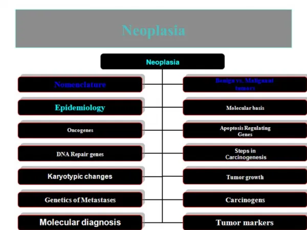
NOMENCLATURE OF NEOPLASMS • Benign tumors • Usually designated by adding “-oma” to cell type • adenoma – benign tumor arising from glandular cells • leiomyoma – benign tumor arising from smooth muscle cells • chondroma – benign tumor arising from chondrocytes • Other benign tumor names • papilloma – has finger-like projections • polyp – projects upward, forming a lump • cystadenoma – has hollow spaces (cysts) inside
Malignant tumors • Carcinomas – arise in epithelial tissue • adenocarcinoma – malignant tumor of glandular cells • squamous cell carcinoma – malignant tumor of squamous cells • Sarcomas – arise in mesenchymal tissue • chondrosarcoma – malignant tumor of chondrocytes • angiosarcoma – malignant tumor of blood vessels • rhabdomyosarcoma – malignant tumor of skeletal muscle cells
Nomenclature Neoplasm Benign Malignant Carcinoma Sarcoma
Nomenclature Neoplasm Benign Malignant adenoma angioma rhabdomyoma Carcinoma Sarcoma
Nomenclature Neoplasm Benign Malignant Carcinoma Sarcoma squamous cell carcinoma adenocarcinoma
Nomenclature Neoplasm Benign Malignant Carcinoma Sarcoma angiosarcoma rhabdomyosarcoma
BIOLOGY OF TUMOR GROWTH • Malignant tumors differ from benign ones by four features that in fact reflect their natural history (expected behavior); these are • 1. Malignant transformation of the target cells • II. Growth rate of the transformed cells • III. Local invasion • IV. Distant metastases
I. Malignant transformation Malignant transformation of target cells is associated with certain microscopic features that are usually used to differentiate benign from malignant neoplasms. *These features are collectively come under the heading of differentiation and anaplasia. *Differentiation signifies "the extent to which neoplastic cells resemble comparable normal cells".
The degree of tumor differentiation is represented by a spectrum according to which neoplasms are divided in to • Well differentiated • Moderately differentiated • Poorly differentiated • Undifferentiated (Anaplastic)
Well-differentiated neoplasms i.e.composed of cells resembling closely comparable normal cells of the tissue of origin. Certain well-differentiated thyroid follicular carcinomas (adenocarcinomas), for example, may form almost normal-appearing follicles, and the cells of some squamous cell carcinomas contain cells that do not differ significantly from normal squamous epithelial cells. Thus, the microscopic diagnosis of malignancy in well-differentiated neoplasms can be quite difficult.
2. Poorly differentiated neoplasms, in contrast,are composed of cells that barely resemble the normal cells of origin.However, some resemblance occurs focally i.e. in some parts of the tumor, thus allowing the tumor to be assigned to a particular cell of origin or differentiation. 3. Moderately differentiated neoplasms occupy a morphological position that lies between well-differentiated & poorly-differentiated tumors. 4. Undifferentiated (anaplastic) neoplasms: anaplasiasignifies total lack of differentiation, and thus anaplastic cells have primitive appearance (unspecialized morphology) that can not be assigned to any of the normal mature cells
Morphologic features of malignant cells include: • Pleomorphism, i.e. variations in the size and shape of the neoplastic cells and their nuclei. In anaplastic cancers, some cancerous cells are many times larger than their neighboring extremely small cancerous cells. 2. Abnormal nuclear morphology: characteristically the nuclei display a. Hyperchromatism,which refers to a deep bluish staining of nuclei; this feature is due to their abnormally high content of DNA. In the routine hematoxyline & eosin stain, the abundant DNA extracts more hematoxyline, and so the malignant nuclei appear deep blue in color.
b. High nuclear-cytoplasmic ratio (high N/C); in malignant neoplasms, the nuclei are disproportionately large for the cell size, and thus the nucleus-to-cytoplasm ratio may approach 1:1 instead of the normal 1:4 or 1:6. c. Variations in nuclear shape and abnormal chromatin clumping and distribution: the nuclear shape is very variable, and the chromatin is coarsely clumped and distributed along the nuclear membrane. Normal nuclei are vesicular i.e. have fine, evenly distributed chromatin granules. d. Large prominent nucleoli are sometimes seen within malignant nuclei.
3. Frequent mitoses including abnormal ones:undifferentiated malignant cells usually possess large number of mitoses, reflecting their high proliferative activity. The presence of mitoses, however, does not necessarily indicate that a tumor is malignant or that the tissue is neoplastic. Many normal tissues exhibit rapid cell turnover & hence their constituent cells show frequent mitoses e.g. the normal bone marrow cells. The adaptive hyperplastic tissue response also shows frequent mitotic figures. More important as a morphologic feature of malignancy is the presence of atypical mitotic figures, e.g. tripolar, quadripolar, or multipolar mitoses (instead of the normal bipolar spindles)
4. Loss of polarity: this means disturbed orientation of the cells. In malignancy, sheets of tumor cells grow in disorganized fashion. Normal epidermis shows normally oriented stratified constituent cells; from below up there are the basal cells followed by spinous cells then granular cells and finally the upper most layer of flattened, keratinized cells. Although differentiated squamous cells carcinoma tend to recapitulate to some extent this arrangement, it is totally lacking in poorly differentiated and undifferentiated examples i.e. there is no longer the orderly architectural stratification seen in the normal skin
5. Other changes a. Formation of tumor giant cells: some of these abnormally large cells possess only a single huge pleomorphic nucleus; others have two or more nuclei. These malignant giant cells are not to be confused with the inflammatory Langhan or foreign body giant cells; the latter are derived from macrophages and contain many small, normal-appearing nuclei. In cancer giant cells, the nuclei show malignant features, for e.g. they are hyperchromatic and large in relation to the cell size.
b. Foci of ischemic necrosis: dividing and growing tumor cells require adequate blood supply for their survival. In many anaplastic tumors, because of the rapid proliferation of the constituent cells and/or scant stromal vascularity, the tumor may overrun the available blood supply. This leads to large areas of ischemic necrosis. The presence of necrotic areas within a malignant tumor is a poor prognostic sign, since it usually reflects an aggressive rapidly growing malignancy.
Dysplasia Having mentioned the features of malignancy, it is pertinent to comment on a closely related condition designated dysplasia. Dysplasia literally means disordered growth. This change is encountered principally in epithelial membranes (e.g. the squamous epithelium of the cervix uteri, skin, and metaplastic bronchial mucosa) and it is characterized by changes that include: 1. Pleomorphism and loss of orientation of the affected cells 2. Frequent presence of hyperchromatic and large nuclei
3. Unusually abundant mitotic figures; although these are almost invariably of normal patterns (bipolar), frequently, however, the mitoses appear in abnormal locationswithin the epithelium, for e.g. in dysplastic stratified squamous epithelium; mitoses are not limited to the basal layers as is the case normally, but appear at all levels including the surface cells. According to the severity & extent of the above changes, dysplasias are graded into mild, moderate and severe; recently dysplasia is also divided into low-grade and high-grade.
When the dysplastic changes are severe and involve the entire thickness of the epithelium, it is considered a pre-invasive neoplasm and is referred to as carcinoma in situ. Once the tumor cells move beyond the normal confines through breaching the limiting basement membrane, the tumor is considered invasive carcinoma. Dysplastic changes are often found adjacent to foci of invasive carcinoma, and in some situations, such as in long-term cigarette smokers, severe epithelial dysplasia of the bronchial epithelium frequently precedes the appearance of cancer. However, dysplasia does not necessarily progress to cancer
. Mild to moderate dysplastic changes of the cervical squamous epithelium i.e. those that do not involve the entire thickness of epithelium may be reversible, and with the removal of the inciting cause, the epithelium may revert to normal. Severe dysplasia (when the full thickness of the epithelium is involved) is much more serious because of its more intimate association with invasive carcinomas.
II. Growth rate of the transformed cells The growth rate of neoplasms (i.e. how rapidly they increase in size) influences not only their clinical outcome but also their response to therapy. Any neoplasm is considered clonal i.e. originating from one (or at most few) initially transformed cells (I-TC). For the tumor to be clinically detectable (at least 1 g in wt), the I-TC and its progeny (collectively referred to as tumor cell population) must undergo at least 30 population doublings
The larger the cancer, the more difficult it becomes to treat and control. Accordingly, diagnostic investigations are needed to detect early cancers & this is the prime goal of screening programs e.g. that of the cervix (Pap smear) & breast (triple assessment i.e. physical examination, mammography and fine needle aspiration cytology).
The growth rate of a tumor is determined by three main factors: 1. The doubling time of tumor cells (length of the cell cycle) 2. The size of the replicative pool. (Replicative pool refers to that part of the tumor made up of exclusively of dividing cells). 3. The rate at which cells leave the growing tumor
III. Local invasion Benign tumors differ from malignant ones by their slow rate of growth growing and as cohesive masses, thus, benign tumors usually (not always) develop a rim of compressed connective tissue called fibrous capsule, which separates them from the native host tissue. An example is fibroadenoma of the breast. This tumor on clinical examination is well-defined and typically mobile mass.
Benign tumors remain confined to the site of origin without having the ability to invade locally ormetastasize to distant sites. In contrast, most malignant tumors are invasive and can be expected to penetrate and destroy the underlying tissues i.e. they are not surrounded by a capsule. Fixation of a breast mass on clinical examination makes it suspicious for malignancy. It is this invasiveness that makes surgical resection of cancers difficult. It is necessary during surgery to remove a margin of apparently normal tissues (margin of safety) adjacent to infiltrative cancer.
One of the prime functions of the pathologist is to indicate, in his report of a surgically excised malignant tumor, of whether the tumor is totally removed (free excision margins) or incompletely excised (positive excision margins). In the latter instance recurrence of the tumor is a strong possibility. Some cancers progress from a pre-invasive stage (carcinoma in situ); this commonly occurs in carcinomas of the skin, breast, and uterine cervix. In situ epithelial cancers display the cytologic features of malignancy but are devoid of invasion outside the encompassing basement membrane.
IV. Metastasis By definition metastases are "tumor implants discontinuous with the primary tumor". Metastasis is the only definitive criterion of malignancybecause benign neoplasms do not metastasize. Almost all cancers can metastasize. The major exceptions are 1. Most malignant gliomas of CNS (gliomas are tumors derived from glial cells) 2. Most Basal cell carcinomas of the skin. Rodent ulceris a clinical descriptive term used for basal cell carcinoma because of their destructive invasiveness. Yet, they do not, as a rule, metastasize.
In general, cancers more likely to metastasize are • The more aggressive and more rapidly growing 2. Those of large size Metastatic spread strongly reduces the possibility of cure. Pathways of Spread Dissemination of cancers may occur through one of three pathways: • Direct seeding of body cavities or surfaces b. Lymphatic spread c. Hematogenous spread
a. Seeding of Body Cavities and Surfaces This may occur whenever a cancer, as a result of progressive invasion, penetrates into a natural "open field." Most often involved is the peritoneal cavity, but any other cavity—pleural, pericardial, subarachnoid, and joint space—may be affected. Peritoneal seeding is particularly characteristic of ovarian carcinomas. b. Lymphatic Spread This is the most common pathway for the initial dissemination of carcinomas, but some sarcomas may also use this route. The pattern of lymph node involvement follows the natural routes of lymphatic drainage.
c. Hematogenous Spread Hematogenous spread is typical of sarcomas but is also seen with carcinomas. Veins, because of their thinner wall, are more readily penetrated than arteries. With venous invasion, the blood-borne cells follow the venous flow draining the site of the neoplasm. The liver and lungs are most frequently involved by metastases. This is because all portal area drainage flows to the liver, and all vena cava blood flows to the lungs.