Introduction to Neoplasia and Tumor Growth
320 likes | 420 Vues
Understand neoplasia basics, cellular differentiation, genetic basis, terminology, and classification of tumors. Explore the gross and microscopic features, causes of cancer, and the impact of environmental factors. Learn key terms and concepts in tumor pathology.

Introduction to Neoplasia and Tumor Growth
E N D
Presentation Transcript
Neoplasia Dr. Bruce F. Burns Anatomical Pathology Ottawa Hospital
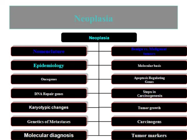
Overview • Characteristics of neoplasms compared to normal tissues • Types of neoplasms • Benign vs malignant • Cellular differentiation • Classification schemes • Genetic basis for neoplasia
What is a “neoplasm”? • Lay term of “tumor” conveys usual connotations – ie a new growth or mass • Definition revolves around these features: • Monoclonal proliferation of cells with specific mutations • Excessive and unregulated growth of these cells, often at the expense of surrounding normal tissue
Terms to know about when discussing neoplasia • Metastasis - spread of a malignant tumor from one site to another via blood or lymph • Benign – typically refers to those tumors incapable of metastasis and having a good clinical outcome (prognosis) • Malignant – those tumors capable of invasive growth and/or metastasis, often fatal if not treated effectively
More terms…. • Parenchyma – these are the tumor cells themselves, usually referring to epithelial cells in organs. • Stroma – connective tissue cells that support the parenchymal cells – not actually tumor cells, but are stimulated to grow by the tumor via growth factors, eg angiogenesis
Cellular differentiation • Tumors are often “graded” as to how closely they resemble the normal parent tissue that they are derived from. • Well-differentiated means the cells are very similar in appearance and architectural arrangement to normal tissue of that organ
Colonic “adenoma” illustrating a “well-differentiated” neoplasm similar to normal colon mucosa
Differentiation • “Poorly-differentiated” refers to tumors that show only minimal resemblance to the normal parent tissue they are derived from. • “Anaplastic” means the tumor shows no obvious similarity to it’s parent tissue, usually associated with aggressive behavior
So what?????? • Differentiation often provides clues as to the clinical aggressiveness of the tumor • Tumors often lose differentiation features over time as they become more “malignant” and as they acquire more cumulative genetic mutations • Differentiation often predicts responsiveness to certain therapies, eg estrogen receptors and Tamoxifen in breast cancers
Gross (macroscopic) features of two breast neoplasms Benign – circumscribed, often encapsulated, pushes normal tissue aside Malignant – infiltrative growth, no capsule, destructive of normal tissues
Classification of neoplasms • Epithelial tumors • Benign forms – adenoma , papilloma • Malignant forms – carcinoma, eg adenocarcinoma, squamous cell carcinoma • Mesenchymal tumors • Benign forms – fibroma, leiomyoma, • Malignant forms – sarcoma, eg fibrosarcoma, leiomyosarcoma
Classification continued • Tumors of lymphocytes are always malignant – called lymphoma • Tumors of melanocytes • Benign – nevus • Malignant - melanoma
Microscopic features of tumors • Loss of normal architectural arrangement –
Microscopic features of tumors • Pleomorphism – variation in size and shape of cells within the neoplasm
Microscopic features of tumors • Mitotic activity - Increased in more malignant tumors and often abnormal in shape
Precursors of neoplasia • Hyperplasia • Metaplasia • Chronic inflammation • dysplasia
Metaplasia, dysplasia, neoplasia • Metaplasia – an adaptive change in differentiation, reversible, no mutations necessary. • Eg- change of esophageal mucosa from squamous to gastric type in the setting of acid reflux (“heartburn”). Better able to withstand the corrosive effects of the acid. • Metaplasia is fertile ground for development of “dysplasia” (disordered growth)
Metaplasia, dysplasia, neoplasia • Dysplasia refers to recognizable morphologic changes in cells that indicate the presence of genetic mutations beginning the development of a neoplasm • Often graded, eg PAP smears for uterine cervical cancer are low and high grade
Causes of Cancer • Most cancer arises as the result of somatic mutations in the genome resulting from: • Chance (ie, we don’t know) • Environmental factors – chemical, radiation, viruses • Ageing • Inherited cancer syndromes- defect in germline DNA
Environmental carcinogens • Chemicals capable of DNA damage • Initiators vs Promoters • Common denominator is “electrophilic intermediates” forming adducts with DNA • Some are direct acting, others are activated in the body, usually in the liver by cytochrome P-450 enzymes
Radiation • Ionizing radiation – x-rays, gamma rays, radioactive materials such as Radon gas – all cause a variety of defects to DNA • UV light (non-ionizing) – primarily sun-exposure and T-T dimerization – skin cancers
Common features of viral carcinogenesis • Oncogenic viruses typically integrate their genomes into host cells and enter a period of “latency” • May be of DNA or RNA type • DNA viruses include EBV, HPV and Hepatitis B virus • RNA viruses include retroviruses like HTLV-1 and indirectly HIV
Viral carcinogenesis • Human papilloma virus (HPV) prototype • Cause warts • Some types have stronger cancer causing associations, esp 16 and 18 with uterine cervix cancer - Pap smears of cervix can detect precursor lesions of infection – Rx • Viral genes interact with human genes concerned with cell division
How does HPV cause cancer? • Gene products of certain sub-type (eg 16 and 18) interfere with normal cellular proteins • Early viral proteins E6 and E7 bind p53 and RB proteins respectively
Other oncogenic viruses • Epstein-Barr virus (EBV) associated with some lymphomas and nasopharyngeal carcinoma • Hepatitis B virus associated with malignant liver tumors