Exploring the Microscopic World with Magnification
230 likes | 270 Vues
Discover the intricate details of microscopic elements with varying levels of magnification, exploring different specimens like teeth, skin, cells, and embryos. Learn about the history and types of microscopes, from compound light to electron microscopes, each offering unique advantages and capabilities. Delve into the fascinating world revealed by these powerful tools.

Exploring the Microscopic World with Magnification
E N D
Presentation Transcript
Tooth with plaque Magnification: 10X
Toothbrush and Tooth Magnification: 75X
Human Finger tip Magnification: 12X
Human Finger tip Magnification: 600X
Human Skin with Bacteria Magnification: 8,000X
Mosquito Magnification: 50X
Human Cells Magnification: 10,000X
Embryonic Hand Magnification 7X 5 weeks Embryonic Hand Magnification 10X 11 weeks Human Embryo Magnification 10X 16 weeks
Stomach Pit with Red Blood Cell and Acid Magnification: 3,000X
Human Brain Magnification: 5X
6 week Human Embryo Magnification: 10X
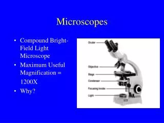
Microscope—tool used to study things too small to be seen by the unaided eye. • History: • Invented by theJansen brothers in 1590. • Improved by Anton Van Leeuwenhoek in the 1600’s. • His first scope had a power of 270X. • He made over 400 different microscopes. • Known as the “Father of Microbiology.” Anton Van Leeuwenhoek
Types of Microscopes • Compound Light Microscope —uses lenses to magnify and light to illuminate. The most powerful can magnify 2000X. • Eyepiece —lens closest to the eye. • Objective lens —lens closest to the object. • Total magnification —the power of the eyepiece times the power of the objective. • Example: 10X x 43X = 430X
Types of Microscopes • Stereoscope —used to view larger objects. The light source is above the object, not below.
Types of Microscopes • Electron Microscope —uses magnets to focus a beam of electrons. • Electrons are negatively charged particles. • Total magnification can be up to 1,000,000X. • Disadvantages—sometimes can not study living organisms; expensive; too powerful • Types: • Transmission Electron Microscope (TEM)—used to study internal parts of a specimen. • Scanning Electron Microscope (SEM)—used to see the surface of whole objects. • Scanning Tunneling Microscope—the most powerful. Invented in 1981. Has a magnification of 100 million times and a resolution of 1/100th the diameter of an atom.
Transmission Electron Microscope • The TEM passes electrons through the object being studied. It can magnify as much as 200,000x but can not be used to observe living tissue.
The TEM gives very detailed pictures of the internal structure of materials, either biological or non-biological. Example: Golgi body of an a animal cell.
Scanning Electron Microscope • The SEM has a magnification range of 15x to 200,000x and a resolution of 5 nanometers. • It produces a detailed, three dimensional black and white image 35x 200x 1000x 35000x
Specimens to be viewed by the SEM must be carefully dried to prevent shriveling and must be made to conduct electricity by coating them with a thin layer of gold in a machine called a sputter coater. Dried specimen Sputter coater
Finally, the specimen is placed in the microscope’s vacuum column through an air-tight door. Air is pumped out and an electron gun emits a beam of high-energy electrons which is focused by a series of magnets.
The Scanning Tunneling Microscope invented by John Wendelken and Joe Carpinelli. The microscope is nicknamed JEOL.
Microscope Diagram Microscope Flipchart Activity Eyepiece Eyepiece Tube Body Revolving Nosepiece Arm Objective lenses Stage Clips Stage Diaphragm Coarse Adjustment Light Fine Adjustment Base