Download
1 / 9
410 likes | 1.76k Vues
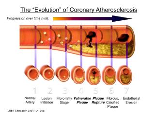
The “Evolution” of Coronary Atherosclerosis. Progression over time (yrs):. Normal Artery. Lesion Initiation. Fibro-fatty Stage. Vulnerable Plaque. Plaque Rupture. Fibrous, Calcified Plaque. Endothelial Erosion. (Libby. Circulation 2001;104: 365).

E N D
The “Evolution” of Coronary Atherosclerosis Progression over time (yrs): Normal Artery Lesion Initiation Fibro-fatty Stage Vulnerable Plaque Plaque Rupture Fibrous, Calcified Plaque Endothelial Erosion (Libby. Circulation 2001;104: 365)
Can the Trajectories of the Natural Histories of Coronary Atherosclerosis Be IdentifiedPrior to Adverse Coronary Events?Opportunities for Intervention Quiescent, Stable plaque no symptoms Fibrotic/ Scarred plaque angina ? Vulnerable, Ruptured Plaque MI, sudden death Snapshot to identify likelihood to develop vulnerability or progression Snapshot to identify vulnerability Snapshot at time of angina or MI
The Effect of Physiologic Shear Stress on Endothelial Structure and Function Physiologic shear stress (~15-50 dynes/cm2) is vasculoprotective: • Enhances endothelial quiescence • - decreases proliferation • Enhances vasodilation • Enhances anti-oxidant status • Enhances anti-coagulant and • anti-thrombotic status (Malek, et al. JAMA 1999; 282:2035)
The Detrimental Effect of Low Shear Stress on Endothelial Structure and Function Low shear stresses and disturbed local flow (< ~ 6 dynes/cm2) are atherogenic: Promotes: • Cell proliferation, migration • Expression of vascular adhesion molecules, cytokines, mitogens • Monocyte recruitment and activation • Procoagulant and prothrombotic state • Local oxidation (Malek, et al. JAMA 1999; 282:2035)
Example of 3-D Reconstruction of Arterial Segment Composite reconstruction of portion of the arterial segment, consisting of outer arterial wall, plaque, and lumen: Original angiogram of a portion of an artery studied Isolated view of reconstructed outer arterial wall: Isolated view of reconstructed lumen: Isolated view of reconstructed atherosclerotic plaque: (Stone, et al. Circulation 2003;108:438)
Coronary Endothelial Shear Stress dynes/cm2 [Artery is displayed as if it were cut and opened longitudinally, as a pathologist would view it.] (Feldman and Stone. Curr Opin Cardiol 2000; 15: 430)
Changes in Native Arteries p<0.001 ESS at Baseline and Vascular Outcomes 6 mo later: Regions of baseline low ESS: • increase in plaque thickness • enlargement of EEM (outward remodeling) p<0.001 p=0.03 Regions of baseline physiologic ESS: • little change in any variable Regions of baseline increased ESS: • increase in lumen radius • increase in EEM radius • decrease in ESS (outward remodeling) (Stone, et al. Circulation 2003;108:438)
Prediction of Areas of Minor ObstructionWhich Are Actively ProgressingIdentification of Limits of Outward Remodeling and Initiation of Lumen Narrowing In-vivo (Confirmation of Glagov Hypothesis) (Feldman, et al 2003, submitted)
New Era of “Preventive” Vascular Approaches:Identification of High-Risk, Minor Obstructions and Application of Focused InterventionsTo Avert Adverse Coronary Events Quiescent, Stable plaque no symptoms Fibrotic/ Scarred plaque angina X X X Vulnerable, Ruptured Plaque MI, sudden death Minor lesion likely to become vulnerable or progress Identification of “vulnerability” Lesion at time of clinical event