Inflammation and white matter diseases
500 likes | 662 Vues
Inflammation and white matter diseases. Infection: an approach based on location:. Infections of the epidural space: epidural empyema Infections of the subdural space: subdural empyema Infections of the subarachnoid space: (lepto)meningitis

Inflammation and white matter diseases
E N D
Presentation Transcript
Infection: an approach based on location: • Infections of the epidural space: epidural empyema • Infections of the subdural space: subdural empyema • Infections of the subarachnoid space: (lepto)meningitis • Infections of the brain itself: abscess and encephalitis
Subarachnoid infection (leptomeningitis) Bacterial infection: purulent meningitis - the arrival of bacteria triggers a classical inflammatory process - it is associated with a generalized cerebral edema: major complication of the acute phase - the process extends in the perivascular spaces ( Virchow-Robin ), and also in the ventricular system via the foramina of Magendie and Luschka
Viral encephalitis: common histological findings - Inflammatory infiltrate in the subarachnoid space, around the blood vessels (perivascular cuffing) and in the parenchyma - microglial proliferation, diffuse or focal - reactive gliosis - neuronal changes: necrosis, neuronophagy - viral inclusions in neurons, astrocytes or oligodendrocytes
Prion diseases Prion diseases TSE's in animals TSE's in man sporadic 85% Scrapie sCJD }14% genetic fCJD ex-Scr acq. by infection GSS CWD FFI TME }1% Kuru BSE iCJD FSE vCJD ex-TSE
Prion diseases • disease where normal prion protein is misfolded (from PrPC to PrPSc) • PrPSc forms aggregates which are a template to transform more PrPC to PrPSc • Original cause can be genetic, infectious or sporadic • Function of PrPC is unknown
Prion diseases • Can affect all grey matter in the brain leading to rapid progressive disease with one or more of: • Dementia (cerebral cortex) • Spasticity (cerebral cortex) • Myoclonus (cerebral cortex) • Cortical blindness • Extrapyramidal signs (basal ganglia, SN) • Ataxia (cerebellum) • Pathology: • Vacuoles • Neuronal loss • Gliosis • Prion protein deposition (sometimes as amyloid plaques)
Againts virus hypothesis • Very resistant agent: • resists dry heat ( > 200ºC) • resists steam autoclaving (up to134ºC, 18 mins.) • resists formaldehyde • resists UV-radiation • resists Gamma-radiation ( > 0.3 MGray) • etc.
Prion theory (Prusiner) • In biochemical separation-infection studies: • Infectivity is not present in a DNA/RNA fraction • Infectivity is present in a protein fraction • In conclusion: • A protein (and that protein only) causes a prion disease
Sporadic Creutzfeldt-Jakob disease diagnosis • Rare disease: 1/106 • Duration: short (months) • Neurological signs and symptoms: Rapidly progressive dementia, myoclonus, ataxia, central visual disturbances, extrapyramidal signs, pyramidal signs, akinetic mutism (variant: chorea, dysesthesias, psychiatric disturbances) • EEG and MRI • Neuropathology • WHO criteria: • Type of CJD (sporadic, genetic, iatrogenic, variant) • Strength of diagnosis (definite, probable, possible)
Familial CJD vs Familial Fatal Insomnia 178Asn-129V-f-CJD 178Asn-129M-FFI
Prion diseases acquired through infection • Kuru • iatrogenic CJD • new variant CJD
Etiology of Kuru • Primary infectious: no fever, no CSF cell raise • Genetic disease: • Pro: in families, in patients who moved out of Fore region. • Con: also in patients who came to live in Fore region, as well in young as in old patients. • Toxic/deficiency: no toxic compound isolated, balanced diet. • Endocanibalism
iCJD number incubation time clinical presentation Intracerebral cornea neurosurgery stereotactic EEG Cerebral surface dura mater* Peripheral blood growth hormone gonadotrophin 4 5 2 174 180 4 17 mo 17 mo 18 mo 6-12 y 12 y 13 y dementia/ataxia dementia/ataxia dementia/ataxia ataxia ataxia ataxia * Mostly Lyodura, now 2 cases with 0.1 N NaOH treated Tutoplast
Clinical history in v-CJD • 65% of cases onset with ‘psychiatric’ complaints: • Behavioural disturbance • Attention deficit • Personality changes • Depression • Later weeks - months: • Dysaesthesia (20 % onset with dysaesth) • Ataxia • Myoclonus/choreo-athetosis • Progressive dementia • No typical EEG • 14-3-3 protein 50% of the cases positive • bilateral Pulvinar sign on MRI
Proposed route of peripheral infection GALT > Spleen B-cell + + Macroph. FDC Macroph. FDC M-cell lymphatics Symp NS Xth CN Gut Spinal cord Brainstem Brain
Protein misfolding to neuronal dysfunction pathogenesis Protein misfolding & accumulation diseases: • Alzheimer’s disease • Parkinson’s disease • Tauopathies • Progressive Supranuclear palsy • Fronto-temporal dementias • Cortico-basal degeneration
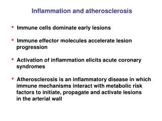
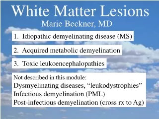
Multiple sclerosis (MS) • Frequently onset in younger individuals (20-40 yoa) • F>M • Exacerbations and remissions (with often only partial remission later in the course) • Locations: central myelin—with symtoms of function loss. • Often initial symptom: visual field defect
Pathogenesis • T cell mediated auto immune disease • Recent hypothesis: venous congestion due to vascular abnormalities of the neck