RF Coil Design Optimization in Cervical Carotid Imaging
260 likes | 468 Vues
RF Coil Design Optimization in Cervical Carotid Imaging. Rock Hadley MIRL Symposium Sundance September 13, 2002. Go Take A Hike!. Available Carotid Coils. 4-channel commercial Neurovascular volume coil. home-built bilateral 4-element carotid Phased-array coil.

RF Coil Design Optimization in Cervical Carotid Imaging
E N D
Presentation Transcript
RF Coil Design Optimization in Cervical Carotid Imaging Rock Hadley MIRL Symposium Sundance September 13, 2002 Go Take A Hike!
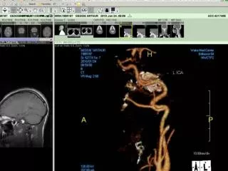
Available Carotid Coils 4-channel commercial Neurovascular volume coil home-built bilateral 4-element carotid Phased-array coil
bilateral 4-element carotid PA coil 300% SNR improvement over anterior neck coil commercial anterior neck coil Dedicated Carotid Coils
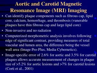
carotid bifurcation circle of Willis relative SNR aortic arch image slice number Coil Comparisons
Magnetic Field of a Circular Loop cross-sectional contour plots coil axis coil signal shadow magnitude (all field components) axial-component (signal component)
Coil Design Issues 12 11 45cm vessel length 25cm vessel length 10 5cm vessel length 1cm vessel length 9 8 7 optimized coil radius (cm) 6 5 4 3 2 1 0 0 1 2 3 4 5 6 7 8 9 10 11 12 13 14 15 vessel depth (cm) long vessels opt coil radius = vessel depth very short vessels opt coil diameter = vessel depth
Simulation Model s = 0, er = 1 (air) coil x surface coil axis depth vessel aortic arch bifurcation circle of Willis s = 0.3, er = 73 (muscle) figure of merit = average SNR along entire vessel (weighting of specific vessel points optional)
Example Simulation • 24 cm vessel length • - 6 cm depth at aortic arch • - 3 cm depth at carotid bifurcation • - 9 cm depth at circle of Willis • Results:
Coil 1 Coil 2 Coil 3 Coil 4 Coil 5 0 flat surface bifurcation 2 4 circle of Willis 6 aortic arch vessel depth (cm) 8 simulated vessel structure 10 12 14 0 5 10 15 20 25 vessel position (cm)
Conclusions • This simulation tool provides a method for the study of optimum coil arrays for various carotid artery anatomy • Number of channels needed for optimal carotid imaging is about 16 – 20 channels
Minimize magnetic and electric coupling between coils to determine proper coil overlap Adapt to non-planer surfaces to more closely match the anatomy of the shoulders, head, and neck Future Work
Coil Properties magnetic field of a circular loop (cross-sectional contour plots) coil axis coil signal shadow magnitude (all field components) axial-component (signal component)
Simulation Results optcoil.results.24fov.639realvessel 1 0.95 0.9 0.85 0.8 max SNR 0.75 0.7 0.65 0.6 0.55 1 2 3 4 5 6 7 8 9 10 # of coils
RF Coil Design Optimization in Cervical Carotid Imaging Rock Hadley MIRL Symposium Sundance September 13, 2002 Go Take A Hike!
Coil Options for Carotid Imaging commercial neurovascular volume coil home-built bilateral 4-element carotid Phased-array coil
C2 C2 C3 C3 C2 C2 C1 C2 C1 C2 L L D1 Gap on back side of copper substrate l /4 triax balun cables Coil layout Dual PA Coil (Hayes et al. Magn Reson Med 18:309-319, 1991)
Methods: Ladder Coil Set UP coil 3 / 5 slabs coil 2 / 3 slabs coil 1 / 4 slabs
Torso PA Inverted Dual PA Chest Dual PA Carotid PA Head Dual PA + GE standard head coil (GE Head) + Commercial Neurovascular PA coils (NVPA)
Develop a coil for MRA screening from the aortic arch to the top of the head Use dedicated coil elements for each section of specific anatomy Ability to use the same coil for high-resolution imaging of suspect regions Purpose