The bodily senses
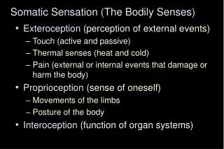
570 likes | 999 Vues
The bodily senses. From Ch. 22 “Principles of Neural Science”, 4 th Ed. Kandel et al. Bodily senses. Bodily senses = somatic sensation Large variety of receptors Distributed throughout the body Sensory information

The bodily senses
E N D
Presentation Transcript
The bodily senses From Ch. 22 “Principles of Neural Science”, 4th Ed. Kandel et al
Bodily senses • Bodily senses = somatic sensation • Large variety of receptors • Distributed throughout the body • Sensory information • Nerves transmit information from the receptors by frequency modulation of electrical signals (action potentials)
Dorsal root ganglion (DRG) • DRG contains the cell bodies of sensory neurons • All somatosensory information from limbs and trunk are transmitted via DRG • Stimulus transmission from sensory receptor to CNS • Primary afferent neuron has two branches to • Periphery • Spinal cord
The sensory receptor • Peripheral receptor • Located at the terminal of the sensory neuron • Molecular specialization that transforms one type of energy into action potentials • Special transducer molecules
Somatic receptor types • Fiber classification • Conduction velocity (skin) or fiber diameter (muscle) • Myelinated or unmyelinated • 4 major modalities (distinct system of receptors and pathways to the brain) • Discriminative touch (size, shape, texture, movement across skin) • Proprioception (joint position) • Nociceptors (tissue damage, inflammation, chemical irritation, pain, itch) • Thermal receptors (warm, cold)
Somatic receptor types 1 2 3 4
Somatic receptor types 1 2 3 4
Somatic receptor types 1 2 3 4
Nerve endings and fiber types • Nerve endings • Bare nerve endings • Thermal and painful (nociception) sensations • Encapsulated nerve endings • Touch and proprioception • Deformation of receptive surface • Large diameter myelinated axons (rapid conduction) • Mechanoreceptors (touch, greatest density in glabrous skin [hairless], finger tips, lips) • Proprioceptors (joint position) • Small diameter myelinated and unmyelinated (slow conduction) • Thermal receptors • Nociceptors
Mechanoreceptors • Specialized organs surrounding the nerve endings • Sensitive to displacement/ deformation • 4 major types in glabrous skin • Superficial location (located below skin ridges) • Meissner corpuscle – in glabrous skin • Rapidly adapting, fluid filled structure, sense deformation of small areas • Merkel disk receptor - in glabrous skin and hairy skin • Slowly adapting, sense sustained pressure, salient bumps, sharp edges • Deep subcutaneous location (less numerous) • Pacinian corpuscle • Similar to Meissner corpuscle, rapid indentation, minute vibration, frictional displacement, small irregularities (edges/ corners) • Ruffini ending • Slowly adapting, links folds in skin at the joints, sense stretch and bending, shape of grasped objects, global properties of objects, wide area of skin
Mechanoreceptors • Deep receptors sense deformation of a wider skin area that extends beyond the overlying ridges • Nerve fibers to superficial layers branch off to several nearby sensory receptors • Nerve fibers in subcutaneous layers only innervate one receptor • 4 types of mechanosensitivity • Gentle touch of skin (well-localized) • Vibration (frequency and amplitude) • Texture (discrimination with fine spatial detail, two-point discrimination) • Shape of objects grasped
Small, well- localized Large, central zone with max sensitivity Receptive field (RF) Direction- specific stretch • Size and structure of RF vary
2-3 mm diameter Fine spatial differences Small, well- localized 10-25 receptors 10 mm diameter Relative sensitivity to pressure Large, central zone with max sensitivity Direction- specific stretch coarse spatial differences Central zone with large continuous surface Directly above receptor Receptive field (RF) • Size and structure of RF vary
Uniform distribution Most numerous receptor types Fine spatial sensitivity Best at finger tips Finger tips are the most densely innervated region of the skin Receptor distribution 300 mechanoreceptive nerve fibers per square centimeter
Two-point discrimination • Two-point discrimination • Min distance as which 2 stimuli can be resolved as distinct • Determine if one or more points are stimulated • Spatial resolution depends on the RF size/ receptor density • Spatial resolution of stimuli varies across the body • Smallest receptive fields in fingers, lips, and tounge
Vibration sense • Vibration is coded by spike trains • Each AP signals one sinusoidal cycle • Vibration frequency is signalled by the AP frequency • Different receptors have different sensitivity • Merkel: 5-15 Hz • Meissner: 20-50 Hz • Pacinian: 60-400 Hz • Detection depends on size of skin indentation and frequency • Detection threshold = tuning threshold = Lowest stimulus intensity that evokes one AP/ cycle • Intensity of vibration depends on the total number of nerve fibers activated Sensory threshold
Adaption and threshold • Slowly adapting (SA) • Constant pressure • Rapidly adapting (RA) (1) Adapting at the beginning and end of stimulus (2) Encode sense of motion of object - Fires when position change (firing rate proportional to speed) - Stops firing when object is at rest • AP/ sec depends on indentation force • Sensory threshold • The minimum stimulus intensity generating an AP • RA’s have lowest touch threshold • Pacinian corpuscles are the most sensitive mechanoreceptor
Constant force Shape and size • P= F/a • At constant force (F), the smaller area (a) stimulated results in bigger pressure (P) => higher firing rate • Strong initial response • Firing rate is proportional to the curvature of each probe
Smaller Receptive field diameter Bigger Higher Spatial resolution Lower Spatial characteristics • Texture, size, and shape are signalled by population of receptors • Periodic firing of groups of receptors signal the spatial characteristic • Active and inactive receptors contribution • The individual receptor is only stimulated by a part of the pattern • The spatial resolution depends on receptor density and type of receptor • Natural stimuli rarely activates a single receptor alone
Example: lifting an object • Lifting and object • Grasp, force increase, object lifted, vertical gravitational pull, force decrease, release • Grasp and release • Meissner c. : contact/ release; increased grasp force • Pacinian c. : transient pressure at start/ stop • Grip force • Merkel disks: continous firing/ proportional with force • Gravitational pull • Ruffini endings: slowly adapting, sense stretch
Somatic receptor types 1 2 3 4
Peak sensitivity Adaption Silenced Thermal receptors 4 thermal sensations: cold, cool, warm, hot • Constant temperature • Adaption • Tonic discharge/ steady rate • Body temperature • Continuously low rate • Cold fibers more active • Most sensitive to changes in temp than constant temp • Warm fibers: • Range, 29-49 deg • Peak/ preferred, 45 deg • Cold fibers: • Range 5-40 deg • Peak/ preferred, 25 deg Encoding of temperature involves comparing the relative activity of different populations
Somatic receptor types 1 2 3 4
Nociceptors • Information about stimuli that can damage tissue are conveyed by nociceptors • Chemicals are released from traumatized tissue • E.g. Substance P, histamine, and bradykinin • 2 overall types: • Nociceptive specific • Wide dynamic range neurons • 3 classes of nociceptors • Mechnical: pinch, punctate, squeeze • Thermal: above 45 deg or below 5 deg Polymodal: mechanical, thermal, chemical Mechanical nociceptor
Somatic receptor types 1 2 3 4 Proprioception: sense of position and movement of one’s own limbs wo. Vision (1) static limb position, (2) limb movement (kinesthesia); in muscle and joints
Afferent fibers • Different size and conduction velocity of axons • Large fibers conduct faster than small/ thin fibers because the internal resistance to current flow is low and nodes of Ranvier are spaced further apart • Myelination sheets increase conduction velocity Compound AP = sum of all activated nerves Spike amplitude is proportional to fiber diameter
Innervations of dorsal roots Dermatomes Important for location of spinal injury
Distinct ascending pathways • Dorsal column-medial lemniscal system • Touch and proprioception from limbs and trunk • Somtatotopically organized from spinal to cortical level • Ascends ipsilateral side • Cross over to contralateral side in medulla • Anterolateral system • Spinal lamina I, IV, V, VII, VII • Pain and temperature from limbs and trunks • Cross over to contralateral side in spinal cord • Somtatotopically organized from spinal to cortical level Contralateral
The perception of pain From Ch. 24 “Principles of Neural Science”, 4th Ed. Kandel et al
Somatic sensations • Somatic sensation = bodily sensation • Pain is a submodality of somatic sensation • Pain and nociception (conscious vs. peripheral) • Pain sensation is the most salient sensation • Pricking • Burning • Aching • Stinging • Soreness • Pain is a warning of actual or potential injury and damage • Pain depends on the psychological state • The same stimulus can result in different responses under similar conditions and in different individuals
Nociceptors • Information about stimuli that can damage tissue are conveyed by nociceptors • Chemicals are released from traumatized tissue • E.g. Substance P, histamine, and bradykinin • 3 classes of nociceptors • Mechnical: pinch, punctate, squeeze • Thermal: above 45 deg or below 5 deg Polymodal: mechanical, thermal, chemical Mechanical nociceptor
Somatic receptor types 1 2 3 4
Afferent fibers • Different size and conduction velocity of axons • Large fibers conduct faster than small/ thin fibers because the internal resistance to current flow is low and nodes of Ranvier are spaced further apart • Myelination sheets increase conduction velocity Compound AP = sum of all activated nerves Spike amplitude is proportional to fiber diameter
DRG Spinal dorsal horn Nociceptive afferents Compound Action Potential First pain: Sharp and pricking, faster A-delta fibers Second pain, burning and dull, slower C-fibers Blocking each nerve blocks the sensation
Spinal dorsal horn neurons • 2 overall types of interneurons: • Nociceptive specific: responds exclusively to noxious stimuli • Wide dynamic range neurons: graded response to non-noxious and noxious stimuli • Lamina I and II • Direct input from mainly A-delta and C fibers. • NS and WDR interneurons • Lamina III and IV • Direct input from A-beta. Nonnoxious input. Topographically organised receptive field • Lamina V • Direct input from A-beta and A-delta. Direct/ indirect from C-fibers. Convergence of visceral afferents. WDR interneurons projecting to brain stem and thalamus. • Lamina VI • Direct input from A-alpha (nonnoxious) from joints and muscle • Lamina VII and VIII • Respond to noxious input. Polysynaptic. Bilateral response
Neurotransmitters • Fast synaptic potentials • Glutamate (amino acid) • Efficient reuptake of amino acids • Range: postsynaptic neurons in vicinity • Slow synaptic potentials • Neuropeptides e.g. Substance P • No reuptake mechanisms • Range: diffusion, many neurons, unlocalized nature of pain • Neuropeptides • Released and increased in persistent pain conditions • Enhances and prolong the actions of glutamate • Application of substance P produces signs of inflammation e.g. heat, redness, and swelling
Chronic pain • Chronic pain appears to serve no useful purpose • Abnormal pain states • Nociceptive and neuropathic • Nociceptive pain • Direct activation of nociceptors • Tissue damage or inflammation • Neuropathic pain • Direct injury to the nerves • Peripheral or central • Burning or electrical sensation
Chronic pain • Spontaneous ongoing pain • Pain of variable intensity and duration • Spontaneous discharges in periphery and centrally • Referred pain • Pain in a location distant from the source. Could be explained by viscero-somatic convergence in lamina V • Hyperalgesia • Increased pain sensitivity • Allodynia • Non-painful input becomes painful e.g. touch on sun burned skin • Allodynia and hyperalgesia only exist during stimulation • Alterations in biochemical properties and excitability of dorsal horn neurons can induce spontaneous pain, hyperalgesia and allodynia
Referred pain • Signals from muscles and viscera can be felt as pain elsewhere • Example: myocardial infarction and angina can be felt in chest and left arm • Mechanism: convergence of afferents muscle/ viscera afferents and somatic afferents. • Convergence on the same projection neurons in the dorsal horn • The brain cannot tell the difference
Hyperalgesia • Peripheral sensitization: • Increased nociceptor sensitivity • Increased spontaneous activity • Central sensitization: • Increased spontaneous activity • Hyperexcitability of spinal dorsal horn neurons • Wind-up: progressive increased response = amplification (depends on glutamate acting on NMDA receptors) • Prolonged after-discharges to afferent input • Expansion of peripheral receptive fields of central neurons • Can be induced by repetitive firing of nociceptive afferents • Primary hyperalgesia • Hyperalgesia in damaged area (within 5-10mm) • Peripheral sensitization • Secondary hyperalgesia: • Hyperalgesia in surrounding undamaged tissue (10-20mm). • Peripheral and central sensitization
Pressure pain thresholds P<0.001 PTS CTR P<0.001 Niddam et al. 2008 Clinical hyperalgesia Myofascial pain patients (PTS) vs. normal controls (CTR) Myofascial trigger points are hyperalgesic contractures in the muscle IMES stimulus-response curves
Pain and the brain • Pain is a subjective conscious experience. Pain does not exist without the brain • CNS inhibitory or facilitatory mechanisms are remarkable efficient in decreasing or amplifying the pain experience • Changes in CNS contributes to chronic pain (reorganization: biochemical, atrophy, functions) • A better understanding of endogenous pain modulatory systems may lead to new mechanism-based therapies and drug targets
Pain and the brain: modulation • Factors that can influence the pain experience • Top-down brain processes • Memories (previous experience) • Emotion • Cognition (attention/ distraction) • Mood (depression, anxiety) • Context (stress, anticipation/ expectation, placebo) • Endogenous pain control systems • Other factors • Genes • Pathological factors (structure, transmitters, receptors, transporters etc.) • Age, gender
Acute vs. chronic pain • Acute pain characteristics • Activation of peripheral receptors under normal conditions • Sensation of pain closely related to the duration of the stimulus • Chronic pain characteristics • Spontaneous ongoing pain • Peripheral sensitization (spontaneous resting activity and hyperexcitable receptors) • Central sensitization (prolonged peripheral input) • Lowered pain threshold (Hyperalgesia) • Non-nociceptive input becomes painful (allodynia) • Functional and structural changes in PNS and CNS • Segmental expansion of receptive fields • De novo synthesis of membrane proteins • Spouting of spinal terminals of afferent fibers • Formation of new synaptic contacts • Altered balance in descending influences
Acute vs. chronic pain • It is important to differentiate between: • Acute and chronic pain states • Different time horizons engage different emotional coping strategies • Chronic pain becomes maladaptive and is highly co-morbid with mood and anxiety disorders • Chronic pain induces CNS changes • Ongoing spontaneous chronic pain vs. perturbations of chronic pain (allodynia/ hyperalgesia) • Passive vs. active coping => medial vs. lateral brain regions?
PFC PFC PFC ACC ACC PCC PPC PPC Insula PCC PCC Thalamus Insula Insula Vermis Amygdala Vermis Neuroimaging of acute pain Muscle pain Cutaneous pain Visceral pain Chen et al. Tooth pain Lu et al., 2004 Lin et al. (preliminary) Niddam et al., 2002
Distinct ascending pathways • Dorsal column-medial lemniscal system • Touch and proprioception from limbs and trunk • Somtatotopically organized from spinal to cortical level • Ascends ipsilateral side • Cross over to contralateral side in medulla • Spinothalamic pathway • Spinal lamina I, V-VII • Pain and temperature from limbs and trunks • Cross over to contralateral side in spinal cord • Somtatotopically organized from spinal to cortical level Contralateral
Ascending pathways • 5 major ascending pathways • Spinothalamic: axons of nociceptive specific and WDR neurons from laminae I and V-VII; contralateral projection, ascends in anterolateral white matter • Spinoreticular:neurons in laminae VII and VIII; anterolateral ascend • Spinomesencephalic: neurons in laminae I and V; anterolateral ascend to PAG, and spinoparabrachial tract to PB, amygdala; pain affect • Cervicothalamic: arises from lateral cervical nucleus; laminae III and IV; some projects via the dorsal column to cuneate and gracile nuclei (large fiber pathway) • Spinohypothalamic: laminae I, V, VIII; autonomic control • Thalamic nuclei • Lateral nuclear group: spinothalamic tract, NS and WDR, laminae I and V, small receptive fields, encoding location of injury • Medial nuclear group: spinoreticulothalamic tract, laminae VII and VIII