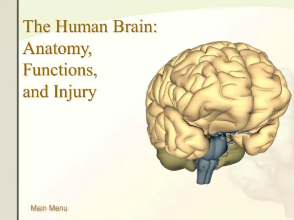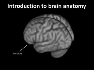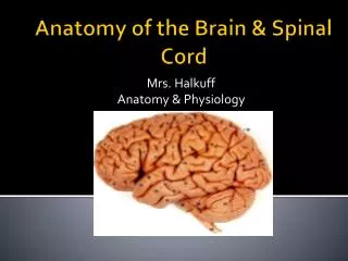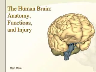
Brain Anatomy
Brain Anatomy. Meninges - 3 layers. Dura Mater Superficial Fuses brain to skull Arachnoid Reduces friction Filled with CSF; shock absorber Pia Mater Very Vascular; needs a lot of oxygen due to high metabolic rate of neurons. Cerebrum Diencephalon Midbrain Pons Medulla Oblongata

Brain Anatomy
E N D
Presentation Transcript
Meninges - 3 layers • Dura Mater • Superficial • Fuses brain to skull • Arachnoid • Reduces friction • Filled with CSF; shock absorber • Pia Mater • Very Vascular; needs a lot of oxygen due to high metabolic rate of neurons
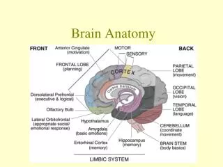
Cerebrum • Diencephalon • Midbrain • Pons • Medulla Oblongata • Cerebellum Gross Anatomy: 6 Regions
Precentral gyrus – Motor Strip Postcentral gyrus – Sensory Strip Central Sulcus – separates frontal from parietal lobe Gyri: elevated ridges Sulci: furrows
Corpus Callosum • Bridge between Right and Left Hemispheres • Enables Right and Left sides to communicate with each other • Problems • “Split Brain” Syndrome
Functions: • Cognition and Memory • Prefrontal Area: involved with intellect, complex learning abilities and personality; plays a role in mood; feelings of frustration and anxiety are formed here • “Gatekeeper” Judgment, critical thinking and reasoning skills • Problems • Relationships between events, memory loss, behavior disorders, Inappropriate social and/or sexual behavior • Prefrontal lobotomy – 1950s Cerebrum: Frontal Lobe
Motor Areas • Function: • Motor Strip: Control voluntary motor function • Premotor Cortex: skill area; controls learned motor skills Precentral gyrus • Broca’s area • Left hemisphere • Directs the muscles of tongue, throat and lips when speaking • Becomes active as we plan to speak • Syntax and grammar rules are remembered
Try This!! Yes the bick. I would say tha the vick daysis nosis or chipickers. Represents problems with Broca’s area!! Only found in the left hemisphere of the frontal lobe Problems will affect our ability to pronounce words, form sentences, speaking becomes a problem
Sensory Areas Located in parietal, temporal and occipital lobes
Parietal Lobe • Primary Somatosensory Cortex • Spatial Discrimination – ability to identify the body region being stimulated • Area is identified by receiving information from skin sensory receptors and proprioceptors in skeletal muscles. • Try This!! • Problems • Inability to locate and recognize body parts; disorientation • Can’t discriminate between different sensory stimuli
Located posterior to Primary Somatosensory Cortex • Major function to analyze different sensory stimuli (temp, pressure • Evaluate what the body is feeling • Try this!! • Different senses are distributed through all lobes Somatosensory Area
Auditory Areas – sound waves are interpreted • Gustatory cortex – perception of taste • Olfactory Cortex – interprets chemical odors • Language • Wernicke’s area – called the speech area • Language comprehension • Understanding jokes • Reading unfamiliar sounds • Problems • Hearing problems • Aphasia – inability to speak Temporal Lobe
Occipital Lobe • Visual Areas • Receives stimuli from eyes • Interprets information from past experiences • Problems • Loss of vision or “seeing stars” • Can’t recognize the object you see
Posterior Association Area • Large region including parietal, temporal and occipital lobes • Plays a role in recognizing faces, patterns, and identifying surroundings • Also includes Wernicke’s area
Diencephalon • Connects to cerebrum • Includes: • Thalamus, • Hypothalamus, • Limbic system • Pineal gland (epithalamus) • Pituitary gland
Thalamus Greek for “Inner room” • Contains relay and processing centers • Relay Station; involved in memory process • Sorts out information, edits • Gateway to cerebrum
Hypothalamus • Controls Body Homeostasis • Autonomic Nervous System • Influences BP • HR (force and rate) • Digestive tract motility • Emotions • Pleasure, fear, rage • Sex Drive • Body temperature regulation • Food intake; hunger • Thirst (water balance) • No blood-brain barrier • Circadian rhythms • Control of Endocrine • (secrete ADH, oxytocin)
Hypothalamus and Pineal Gland Problems with hypothalamus Pineal Gland Part of epithalamus Secretes hormone melatonin Helps regulate Sleep-Wake Cycle • Problems • Hormonal Imbalances • Hypothermia • Diabetes • Obesity • Sleep Disturbances • Dehydration
Hypothalamus is heart of Limbic System: Emotional Brain • Contains Amygdala • Recognizes angry or fearful facial expressions • Contains Hippocampus • Involved with learning, long-term memory and storage • Problems • H.M. Case Study STM to LTM • Had difficulty remembering anything after his surgery • Was able to learn new motor skills, despite not being able to remember learning them Limbic System “Ring”
Link between NS and Endocrine system • Produces GH and TSH • Posterior part of gland is a hormone storage area Pituitary Gland Pituitary Gland ->
Brain Stem: Midbrain, Pons, Medulla Oblongata • Primitive Brain • Pathway between lower brain and spinal cord and lower brain and higher brain functions
Contains 2 pairs of sensory nuclei (Colliculi); Auditory and Visual Reflex Centers I.e. rxns to flashlight or loud noises • Motor nuclei for 2 cranial nerves (III, IV) involved in eye movements • III Oculomotor – eye movement • IV Trochlear – rotates eye up and down • Cerebral Peduncles – descending bundles of motor nerve fibers – connect to cerebellum • RAS center begins here; Filters out repetitive sensory stimuli. (99% of all stimuli is ignored) Midbrain
Colliculi of Midbrain • Corpus Quadrigemini • Superior Colliculi • Visual Reflex Centers • Follow movement with eye • Associated with Cranial nerve III • Inferior Colliculi • Auditory Reflex • Startle Reflex Midbrain
Bridge: Connects cerebellum to brain stem; cerebrum and S. cord Relay Center Cranial Nerves (V-VIII) are attached here Respiratory Center – Involuntary Control of pace and depth of breathing Problems Hyperventilation Pons Bridge
Connects Brain to S. cord; relays info to Thalamus Contains major centers for Autonomic Regulation such as HR, Bp, respiration and digestive activities Cardiac Center – adjusts force and rate of heart beat Vasomotor Center – regulates BP Respiratory Center – controls rate and depth of breathing with N. Fdbk loop in pons. Controls otherpleasant body Activities: vomit, hiccupps, cough, sneeze, swallow, and gag Again no blood-brain barrier! Medulla Oblongata
Coordination; fine tunes voluntary and involuntary movement (Sports) • Receives stimuli from proprioceptors – evaluate body position • Maintains balance and posture • Imbalances • Ataxia; Lack of coordination • Tremors • Alcohol – affects motor skills; reaction time • Easily passes through blood-brain barrier Cerebellum