
Cellular Structure
E N D
Presentation Transcript
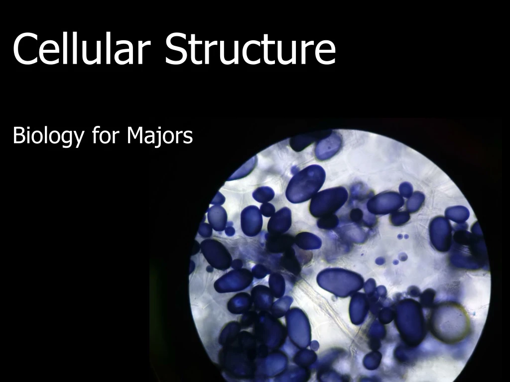
Cellular Structure Biology for Majors
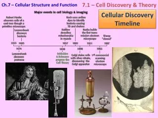
Microscopy A microscope is an instrument that magnifies an object • Light microscope uses light to illuminate a specimen • Electron microscope uses a beam of electrons to illuminate a specimen. It has higher magnification, higher resolution, and more detail
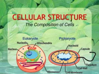
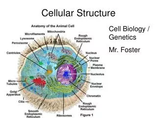
Studying Cells Most cells are too small to see without the aid of a microscope. Within cells there are numerous structures that also need microscopes, especially electron microscopes. In eukaryotic cells, numerous organelles have specialized structures and functions.
Cytoplasm • Different in eukaryotes and prokaryotes. • In eukaryotic cells, which have a nucleus, the cytoplasm is everything between the plasma membrane and the nuclear envelope. • In prokaryotes, which lack a nucleus, cytoplasm simply means everything found inside the plasma membrane. • One major component is the gel-like cytosol, a water-based solution that contains ions, small molecules, and macromolecules. • The cytoskeleton, a network of fibers that supports the cell and gives it shape, is found in the cytoplasm. • Many metabolic reactions, including protein synthesis, take place in this part of the cell.
Nucleus The nucleushouses the cell’s DNA and directs the synthesis of ribosomes and proteins.
Chromosomes and Chromatin Image (a) shows various levels of the organization of chromatin; (b) shows paired chromosomes.
Nucleolus A darkly staining area within the nucleus called the nucleolus aggregates the ribosomal RNA with associated proteins to assemble the ribosomal subunits that are then transported out through the pores in the nuclear envelope to the cytoplasm.
Ribosome Ribosomes are made up of a large subunit (top) and a small subunit (bottom). During protein synthesis, ribosomes assemble amino acids into proteins. All cells have ribosomes.
Mitochondria • During cellular respiration mitochondria make adenosine triphosphate (ATP), the cell’s main energy-carrying molecule. • They have their own DNA and ribosomes.
Peroxisomes Peroxisomes are small, round organelles enclosed by single membranes. They carry out oxidation reactions that break down fatty acids and amino acids. They also detoxify many poisons that may enter the body. Glyoxysomes, which are specialized peroxisomes in plants, are responsible for converting stored fats into sugars.
The Endomembrane System Modifies packages, and transports lipids and proteins.It includes: • the nuclear envelope • lysosomes • vesicles • the endoplasmic reticulum • Golgi apparatus • the plasma membrane.
Endoplasmic Reticulum A series of interconnected membranous sacs and tubules • the rough ER modifies proteins (ribosomes on its surface give it a studded look) • the smooth ER synthesis of carbohydrates, lipids, and steroid hormones; detoxification of medications and poisons; and storage of calcium ions.
Golgi Apparatus Sorting, tagging, packaging, and distribution of lipids and proteins takes place in the Golgi apparatus, a series of flattened membranes. In plant cells the Golgi apparatus also makes polysaccharides.
Vesicles Vesicles are membrane-bound sacs that function in storage and transport. Vesicles can fuse with the plasma membrane to release their contents outside the cell. Vesicles can also fuse with other organelles within the cell.
Unique Features of Animal Cells: The Centrosome The centrosomeconsists of two centrioles (right). They appear to have some role in pulling the duplicated chromosomes to opposite ends of the dividing cell.
Unique Features of Animal Cells: Lysosomes In addition to their role as the digestive component and organelle-recycling facility of animal cells, lysosomes are considered to be parts of the endomembrane system. Lysosomes also use their hydrolytic enzymes to destroy pathogens that enter the cell.
Unique Features of Plant Cells: Chloroplasts • Chloroplasts are organelles that carry out photosynthesis to make food from sunlight. • have their own DNA and ribosomes • contain a green pigment called chlorophyll
Unique Features of Plant Cells: Vacuoles Vacuoles are membrane-bound sacs that function in storage and transport. The membrane of a vacuole does not fuse with the membranes of other cellular components. Additionally, some agents such as enzymes within plant vacuoles break down macromolecules. Thecentral vacuole plays a key role in regulating a plant cell’s concentration of water in changing environmental conditions.
The Cytoskeleton The cytoskeleton is the network of protein fibers that help maintain the shape of the cell, secure some organelles in specific positions, allow cytoplasm and vesicles to move within the cell, and enable cells within multicellular organisms to move.
Types of Fibers in the Cytoskeleton Microfilaments thicken the cortex around the inner edge of a cell; like rubber bands, they resist tension. Microtubulesare found in the interior of the cell where they maintain cell shape by resisting compressive forces. Intermediate filaments are found throughout the cell and hold organelles in place.
The Cytoskeleton: Microfilaments Microfilaments function in cellular movement. They provide some rigidity and shape to the cell. They can disassemble and reform quickly, thus enabling a cell to change its shape and move. Important in immune and muscle cells.
The Cytoskeleton: Intermediate Filaments Intermediate filaments consist of several intertwined strands of fibrous proteins. They bear tension, maintaining the shape of the cell, and anchor the nucleus and other organelles in place.
The Cytoskeleton: Microtubules Microtubules help the cell resist compression, provide a track along which vesicles move through the cell, and pull replicated chromosomes to opposite ends of a dividing cell. They can dissolve and reform quickly.
Flagella and Cilia Hair-like structures that extend from the plasma membrane and are used to move an entire cell. Flagella are longer and cells have 0-3 of them. Cilia are shorter and usually cover the whole outside of the plasma membrane. They may also be used to move substances such as particulate matter in the respiratory tract.
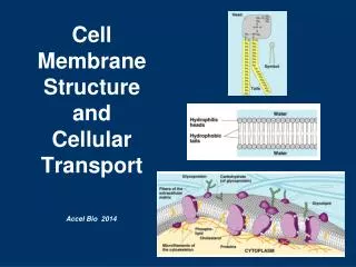
Plasma Membrane Both prokaryotic and eukaryotic cells have a plasma membrane, a phospholipid bilayer with embedded proteins, that separates the internal contents of the cell from its surrounding environment.
Microvilli The plasma membranes of cells that specialize in absorption are folded into fingerlike projections called microvilli.
Plant Cell Junctions Plant cells are connected and communicate with each other via plasmodesmata, channels that pass between cell walls of adjacent plant cells
Animal Cell Junctions Animal cells communicate via their extracellular matrices and are connected to each other via tight junctions, desmosomes, and gap junctions.
The Extracellular Matrix The extracellular matrix holds cells together to form tissues and enables cells within a tissue to communicate. When protein receptors on the surface of the plasma membrane of an animal cell bind to a substance in the extracellular matrix, a chain of reactions begins that changes activities taking place within the cell.
Animal Cell Junctions: Gap Junctions Gap junctions are channels between adjacent cells that allow for the transport of ions, nutrients, and other substances that enable cells to communicate. They are important in cardiac muscle.
Animal Cell Junctions: Tight Junction A tight junction is a watertight seal between two adjacent cells. • found in epithelial tissues • prevents leaking
Animal Cell Junctions: Desmosomes Desmosomes join two adjacent cells together and maintain the cells in a sheet-like formation in organs and tissues that stretch, like the skin, heart, and muscles.
Cell Walls The cell wall is a rigid covering that protects the cell, provides structural support, and gives shape to the cell. Fungal and protistan cells also have cell walls, as do some prokaryotic cells. While the chief component of prokaryotic cell walls is peptidoglycan, the major organic molecule in the plant cell wall is cellulose (below).
Practice Question In this unit we considered the different cell components of cells and focused on the differences between prokaryotic and eukaryotic cells and the differences between plant and animal cells. Which of the cell components (listed on the last two slides) are found in fungi and protists?
Quick Review • Why and how are light microscopes and electron microscopes used in biology? • What is the structure and function of membrane-bound organelles found in eukaryotic cells? • What are the components of the cytoskeleton? • What are cell surface specializations?