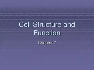
Ch.7 – Cellular Structure and Function
Ch.7 – Cellular Structure and Function. 7.1 – Cell Discovery & Theory. Cellular Discovery Timeline. Ch.7 – Cellular Structure and Function. 7.1 – Cell Discovery & Theory. Why are cells so small??. Surface Area to Volume Ratio.

Ch.7 – Cellular Structure and Function
E N D
Presentation Transcript
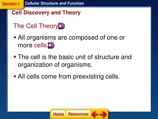
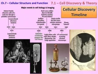
Ch.7 – Cellular Structure and Function 7.1 – Cell Discovery & Theory Cellular Discovery Timeline
Ch.7 – Cellular Structure and Function 7.1 – Cell Discovery & Theory Why are cells so small?? Surface Area to Volume Ratio http://docs.google.com/viewer?a=v&q=cache:AYUlnn-3cIwJ:colgurchemistry.com/Sc10/Sc10BIOLOGY/PDFS/Sc10BiologyAct10SurfaceAreaVolumePDF.pdf+cell+surface+area+activity&hl=en&gl=us&pid=bl&srcid=ADGEESiazsuguvEmmp1KXAJhcYFOzsFfua200s75SY7fYU5wnHpcPGBcRmzf_n9jS4yQEGo9GBk2YBo9LoJq_R0eiuWdLpdm7prBcGXNUv-6IqiZRsgcrHj029YpizX-PEpppWpnuyNR&sig=AHIEtbSM1_pw7JehC0bArlwBXc_4wXlJfQ
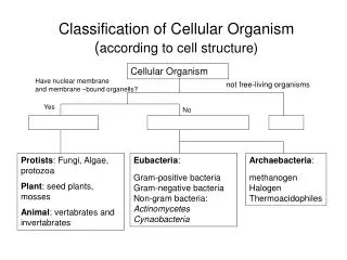
Ch.7 – Cellular Structure and Function 7.1 – Cell Discovery & Theory Cell Types Eukaryotes Prokaryotes Plant Animal Bacteria
Ch.7 – Cellular Structure and Function 7.1 – Cell Discovery & Theory Prokaryotes • Are much more diverse in both habitat and metabolism • Are usually single-celled. Differentiation into different cell types almost never occurs. • Have no separate nucleus. • The cell is surrounded by a membrane, and cell wall but there are no internal membranes. (Few organelles)
Ch.7 – Cellular Structure and Function 7.1 – Cell Discovery & Theory Eukaryotes Cells with a nucleus and membrane-bound organelles Plant Cells Animal Cells Cell Wall Chloroplasts Large Vacuole More geometric shape Centrioles Lysosomes More spherical shape
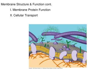
Ch.7 – Cellular Structure and Function 7.2 – The Plasma Membrane The Cell Membrane Organelle/Membrane interactive animation Cell Membranes are Selectively Permeable and are primarily made up of phospholipids
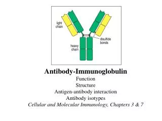
Ch.7 – Cellular Structure and Function 7.2 – The Plasma Membrane The Cell Membrane Other Parts Cholesterol Makes the lipid bi-layer less fluid Glycolipids & Glycoproteins Carbohydrates attached to proteins or lipids and used for cell recognition (Antigens) Proteins Used to transport molecules across the cell membrane (among other uses)
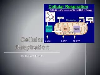
Ch.7 – Cellular Structure and Function 7.4 – Cellular Transport Cellular Transport So, just how does stuff enter and exit cells? H2O, CO2 and small ions freelycross the cell membrane following the natural laws of diffusion Molecules move from areas of high concentration to areas of low concentration This is called… moving with or down the concentration gradient Diffusion continues until Equilibrium is reached
Ch.7 – Cellular Structure and Function 7.4 – Cellular Transport Water is special…. The diffusion of water, across a membrane, down the concentration gradient is called OSMOSIS A simple rule of osmosis is that SOLUTES SUCK Solutes, concentrated inside or outside of the cell, will draw the water in that direction
Ch.7 – Cellular Structure and Function 7.4 – Cellular Transport Hypertonic Isotonic Hypotonic 20% 10% 5% 10% 10% 10%
Ch.7 – Cellular Structure and Function 7.4 – Cellular Transport What will happen to these cells? Hypertonic Isotonic Hypotonic
Ch.7 – Cellular Structure and Function 7.4 – Cellular Transport The artificial cell is permeable to water and monosaccharides only • Draw this diagram • Draw a solid arrow to indicate which direction the solutes will move. • Is the solution Hyper-, Hypo-, or Isotonic Glucose H2O Fructose Hypotonic 4. Draw a dashed arrow to show the movement of water 5. Will the artificial cell become more flaccid, turgid, or stay the same? Turgid
Ch.7 – Cellular Structure and Function 7.4 – Cellular Transport Transport proteins allow Facilitated Diffusion to occur • They create a tunnel that allows specific substances to cross the bilayer down the concentration gradient • They are specific to one substance • They are used for large molecules or to increase the diffusion rate of important substances (water & glucose)
Ch.7 – Cellular Structure and Function 7.4 – Cellular Transport Passive Transport Active Transport Diffusion Osmosis Facilitated Diffusion • Requires ENERGY • Substances move AGAINST the concentration gradient • Requires NO ENERGY • Substances move WITH the concentration gradient Protein Pumps Endocytosis Exocytosis
Ch.7 – Cellular Structure and Function 7.4 – Cellular Transport Protein Pumps Carrier Proteins that use ATP (energy) to move molecules into or out of the cell against the concentration gradient OUT IN Example: Sodium/Potassium Pump Na/K pump animation
Ch.7 – Cellular Structure and Function 7.4 – Cellular Transport Endocytosis Cells take in molecules by forming vesicles at the cell membrane 2 Types Pinocytosis Phagocytosis The cell “gulps” liquid material The cell engulfs solid material
Ch.7 – Cellular Structure and Function 7.4 – Cellular Transport Exocytosis Cells excrete molecules by fusing vesicles to the cell membrane Endo- / Exocytosis Animation
Ch.7 – Cellular Structure and Function 7.4 – Cellular Transport Identify the following: 1. Protein Pump 2. Diffusion 3. Facilitated diffusion