Download
1 / 40
400 likes | 1.14k Vues
Membrane Structure & Function cont. I. Membrane Protein Function II. Cellular Transport. Integral proteins span lipid bilayer called transmembrane proteins hydrophobic regions consist of one or more stretches of nonpolar amino acids often coiled into alpha helices

E N D
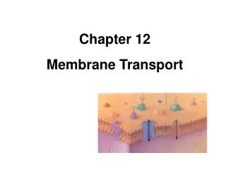
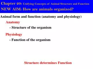
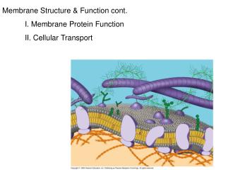
Membrane Structure & Function cont. I. Membrane Protein Function II. Cellular Transport
Integral proteins • span lipid bilayer • called transmembrane proteins • hydrophobic regions consist of one or more stretches of nonpolar amino acids • often coiled into alpha helices • Visualize and draw membrane with transmembrane protein containing 2 helices
EXTRACELLULAR SIDE N-terminus LE 7-8 C-terminus CYTOPLASMIC SIDE a Helix
Six major functions of membrane proteins: • Transport • Enzymatic activity • Signal transduction • Cell-cell recognition • Intercellular joining • Attachment to the cytoskeleton and extracellular matrix (ECM)
LE 7-9a Signal Enzymes Receptor ATP Enzymatic activity Transport Signal transduction
LE 7-9b Glyco- protein Attachment to the cytoskeleton and extra- cellular matrix (ECM) Cell-cell recognition Intercellular joining
The Role of MembraneCarbohydrates in Cell-Cell Recognition • Cells recognize each other by binding to surface molecules, often carbohydrates, on the plasma membrane • Carbohydrates covalently bonded to lipids (glycolipids) or more often to proteins (glycoproteins) • Much variability of extracellular carbohydrates among species, individuals, cell types in an individual • Example of Pneumococcus
Synthesis and Sidedness of Membranes • Membranes distinct inside and outside faces • Plasma membrane is added to by vesicles from ER & Golgi. • Secreted and integral membrane proteins, lipids and associated carbohydrates transported to membrane by these vesicles.
ER Transmembrane glycoproteins LE 7-10 Secretory protein Glycolipid Golgi apparatus Vesicle Plasma membrane: Cytoplasmic face Extracellular face Transmembrane glycoprotein Secreted protein Plasma membrane:
Transport across cellular membranes To exchange materials with surroundings in part to take in nutrients and give off waste Exchange(or transport) regulated: selective permeability
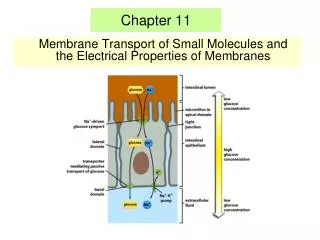
Structure Dictates Membrane Permeability • Hydrophobic (nonpolar) molecules cross membrane rapidly • e.g., hydrocarbons, oxygen, CO2 can dissolve in the lipid bilayer and pass through the membrane rapidly • Polar molecules cross slowly • e.g. sugars, charged proteins, water
How do hydrophilic substances cross membranes? With Help! • Transport proteins • Some create hydrophilic channels across membranes for polar molecules or ions to pass through Example: Aquaporin water channel protein
Carrier proteins binds solutes & change the shape of carrier help to facilitate passage across membrane highly specific for transported solutes Examples: glucose transporter is a carrier protein for glucose only
Passive Transport: Diffusion LE 7-11a Molecules of dye Membrane (cross section) WATER Net diffusion Net diffusion Equilibrium Diffusion of one solute
Substances diffuse down their concentration gradient High to low • Substances reach dynamic equilibrium • No work (no added energy) required
LE 7-11b Net diffusion Net diffusion Equilibrium Equilibrium Net diffusion Net diffusion Diffusion of two solutes
Effects of Osmosis on Water Balance • Osmosis • diffusion of water across a selectively permeable membrane • Diffuses across a membrane from the region of lower solute (such as an ion) concentration to the region of higher solute concentration • The direction of osmosis is determined only by a difference in total solute concentration
Lower concentration of solute (sugar) Higher concentration of sugar Same concentration of sugar LE 7-12 H2O Selectively permeable mem- brane: sugar mole- cules cannot pass through pores, but water molecules can Osmosis
Water Balance of Cells Without Walls • Tonicity • ability of a solution to cause a cell to gain or lose water • Isotonic solution solute concentration is equal inside and outside the cell --> no net water movement cell remains same size
Hypertonic solution external solute concentration is greater than that inside the cell-->cell loses water
Hypotonic solution external solute concentration is less than that inside the cell--> cell gains water May expand enough to burst!
Isotonic solution Hypertonic solution Hypotonic solution Animal cell LE 7-13 H2O H2O H2O H2O Shriveled Normal Lysed Plant cell H2O H2O H2O H2O Flaccid Plasmolyzed Turgid (normal)
Water Balance of Cells with Walls vs No Walls • Cell walls help maintain water balance • Plant cell in hypotonic solution swells -->turgid (firm) • Animal cell? • Plant cell and its surroundings isotonic--> no net water movemen; the cell becomes flaccid (limp), and the plant may wilt • Animal cell? • In hypertonic environment, plant cells lose water--> membrane pulls away from the wall: plasmolysis • Lethal • Animal cell?
Passive Transport Aided by Proteins • Facilitated diffusion • transport proteins speed movement of molecules across the plasma membrane • Channel proteins • Carrier proteins
LE 7-15a EXTRACELLULAR FLUID Solute Channel protein CYTOPLASM
LE 7-15b Carrier protein Solute
Active transportuses energy to move solutes against their gradients • Requires energy, usually ATP • Performed by specific membrane proteins • Example • sodium-potassium pump
EXTRACELLULAR FLUID [Na+] high [K+] low Na+ Na+ Na+ Na+ Na+ LE 7-16 Na+ Na+ Na+ ATP [Na+] low [K+] high P P Na+ CYTOPLASM ADP Phosphorylation causes the protein to change its conformation, expelling Na+ to the outside. Na+ binding stimulates phosphorylation by ATP. Cytoplasmic Na+ bonds to the sodium-potassium pump K+ K+ K+ K+ K+ P K+ P K+ is released and Na+ sites are receptive again; the cycle repeats. Loss of the phosphate restores the protein’s original conformation. Extracellular K+ binds to the protein, triggering release of the phosphate group.
Passive transport Active transport LE 7-17 ATP Facilitated diffusion Diffusion
Electrogenic pumps • is a transport protein that generates a voltage across a membrane--> opposite charges across membrane (membrane potential) • Example: In animals, Na-K pump • In plant fungi and bacteria, proton pump • Requires ATP (active transport)
– EXTRACELLULAR FLUID + LE 7-18 – ATP + H+ H+ Proton pump H+ – + H+ H+ – + CYTOPLASM H+ – +
Cotransport Coupled Transport by a Membrane Protein When active transport of one solute indirectly drives transport of another Example Plants commonly use the proton gradient generated by proton pumps to drive transport of nutrients into the cell
– + H+ ATP H+ – LE 7-19 + Proton pump H+ H+ – + H+ – + H+ Diffusion of H+ Sucrose-H+ cotransporter H+ – + – + Sucrose
How do large molecules move in and out of cells? • Small molecules and water enter or leave the cell through the lipid bilayer or by transport proteins • Large molecules, such as polysaccharides and proteins, cross the membrane via vesicles
Exocytosis • Transport vesicles with cargo migrate to the membrane, fuse with it, and are release contents • Example: • Many secretory cells use exocytosis to export their products • Pancreatic cells (beta-cells) secrete insulin
ER Transmembrane glycoproteins LE 7-10 Secretory protein Glycolipid Golgi apparatus Vesicle Plasma membrane: Cytoplasmic face Extracellular face Transmembrane glycoprotein Secreted protein Plasma membrane:
Endocytosis • Cell takes in macromolecules by forming vesicles at the plasma membrane • Reversal of exocytosis, involving different proteins
Three types of endocytosis • Phagocytosis (“cellular eating”): Cell engulfs particle in a vacuole • Pinocytosis (“cellular drinking”): Cell creates vesicle around fluid • Receptor-mediated endocytosis: Binding of ligands to receptors triggers vesicle formation
RECEPTOR-MEDIATED ENDOCYTOSIS Coat protein LE 7-20c Coated vesicle Receptor Coated pit Ligand A coated pit and a coated vesicle formed during receptor- mediated endocytosis (TEMs). Coat protein Plasma membrane 0.25 µm