
ARGUS® II RETINAL PROSTHESIS
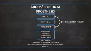
ARGUS® II RETINAL PROSTHESIS. ABSTRACT. MAIN MENU. CLICK A BUTTON TO BEGIN!. Haley Fowler Ed Skolnick Pranali Tambe Terrance White. Instructions. Duke University Department of Biomedical Engineering BME 525: Biomedical Materials and Artificial Organs Fall 2013. ABSTRACT.

ARGUS® II RETINAL PROSTHESIS
E N D
Presentation Transcript
ARGUS® II RETINAL PROSTHESIS ABSTRACT MAIN MENU CLICK A BUTTON TO BEGIN! Haley Fowler Ed Skolnick PranaliTambe Terrance White Instructions Duke University Department of Biomedical Engineering BME 525: Biomedical Materials and Artificial Organs Fall 2013
ABSTRACT Second Sight’s Argus II Retinal Prosthesis is a revolutionary device intended to help restore partial vision to patients suffering from profound blindness caused by a degenerative disease known as Retinal Pigmentosa. This device is the first of its kind and was the first retinal implant to receive Europe’s CE Mark and US FDA approval. These prestigious accomplishments were achieved after 25 years of research and development and a successful 30-patient clinical trial. The Argus II is a device composed of three separate components: a retinal implant, a pair of eye glasses, and a video processing unit. Although all three parts are essential for the device’s proper function, the retinal implant is the primary focus of this module. This module can be used to explore the ins and outs of Argus II’s retinal implant. To begin this module please click one of the buttons to the left. TITLE PAGE INSTRUCTIONS MAIN MENU [14] [10]
INSTRUCTIONS Clicking this will return you to the instructions page. The button on the left below is the standard button used throughout the presentation. Click these buttons to get details about the topic written specifically on the button. Once you have clicked a button and are on that page, the button will appear like the button on the right below. INSTRUCTIONS TITLE PAGE These buttons represent your pathway placement within the module. At any time within the presentation click on any of these buttons to get to a given section. BUTTON BUTTON CLICK HERE TO EXPLORE!! Click the CURVED arrow in order to go back to your last viewed page. Click the check mark to finish a quiz and go back to the main menu. Look for words that are hyperlinked. Click on them to learn more information. HYPERLINKED Click the LEFT and RIGHT arrow to get to the beginning of each subcategory. When an arrow is gray the option is unavailable.
MAIN MENU ARGUS II RETINAL PROSTHESIS [14] [13] IMPLANT & MATERIAL DESIGN BACKGROUND TITLE PAGE [16] [15] Instructions References ACKNOWLEDGEMENTS ABSTRACT DISEASE STATE & PATIENTS SURGICAL PROCEDURE & PERFORMANCE
Learn about anatomy & physiology of the eye, eye implants & the basics of the Argus II! MAIN MENU ARGUS II RETINAL PROSTHESIS [14] [13] IMPLANT & MATERIAL DESIGN BACKGROUND TITLE PAGE [16] [15] Instructions References ACKNOWLEDGEMENTS ABSTRACT DISEASE STATE & PATIENTS SURGICAL PROCEDURE & PERFORMANCE
MAIN MENU ARGUS II RETINAL PROSTHESIS [14] [13] IMPLANT & MATERIAL DESIGN BACKGROUND TITLE PAGE [16] [15] Instructions References Learn about the causes, symptoms, diagnosis, & treatment of RP ACKNOWLEDGEMENTS ABSTRACT DISEASE STATE & PATIENTS SURGICAL PROCEDURE & PERFORMANCE
Learn about the implant design & material selection! MAIN MENU ARGUS II RETINAL PROSTHESIS [14] [13] IMPLANT & MATERIAL DESIGN BACKGROUND TITLE PAGE [16] [15] Instructions References ACKNOWLEDGEMENTS ABSTRACT DISEASE STATE & PATIENTS SURGICAL PROCEDURE & PERFORMANCE
MAIN MENU ARGUS II RETINAL PROSTHESIS [14] [13] IMPLANT & MATERIAL DESIGN BACKGROUND TITLE PAGE [16] [15] Instructions References Learn about the implantation & the performance of the device! ACKNOWLEDGEMENTS ABSTRACT DISEASE STATE & PATIENTS SURGICAL PROCEDURE & PERFORMANCE
BACKGROUND: OVERVIEW BACKGROUND MAIN MENU CLICK TO TAKE “BACKGROUND” QUIZ ANATOMY OF THE EYE 2 PHYSIOLOGY OF THE EYE 1 4 3 HISTORY WHAT IS ARGUS II [13]
DISEASE STATE & PATIENTS: OVERVIEW DISEASE STATE & PATIENTS MAIN MENU CLICK TO TAKE “DISEASE STATE & PATIENTS” QUIZ 1 2 OVERVIEW SYMPTOMS 5 3 CAUSES DIAGNOSIS [16] TREATMENT 4
IMPLANT & MATERIAL DESIGN: OVERVIEW IMPLANT & MATERIAL DESIGN MAIN MENU CLICK TO TAKE “IMPLANT & MATERIAL DESIGN” QUIZ 1 2 STERILIZATION PACKAGING 4 3 IMPLANT MATERIALS IMPLANT DESIGN [14]
SURGICAL PROCEDURE & PERFORMANCE: OVERVIEW SURGICAL PROCEDURE & PERFORMANCE MAIN MENU CLICK TO TAKE “SURGICAL PROCEDURE & PERFORMANCE” QUIZ 2 PERFORMANCE IMPLANTATION PROCEDURE 1 4 3 POST-IMPLANT CARE FUTURE RESEARCH [15]
ANATOMY OF THE EYE BACKGROUND ANATOMY OF THE EYE MAIN MENU OPTIC NERVE HEAD RETINA VESSELS SCLERA CORNEA CHOROID Click each word to learn more about that part. PUPIL LENS IRIS CILIARY BODY [H1] CLICK HERE TO LEARN MORE ABOUT RETINAL ANATOMY!! OPTIC NERVE RETINA Click NEXT to learn more about “Physiology of the Eye”
OPTIC NERVE HEAD BACKGROUND ANATOMY OF THE EYE MAIN MENU Optic nerve head: The location where ganglion cell axons exit the eye to form the optic nerve.This is also the location of the physiological blind spot. OPTIC NERVE HEAD RETINA VESSELS SCLERA [H80] CORNEA CHOROID PUPIL LENS IRIS Optic Nerve Head CILIARY BODY [H1] CLICK HERE TO LEARN MORE ABOUT RETINAL ANATOMY!! OPTIC NERVE RETINA Click NEXT to learn more about “Physiology of the Eye”
RETINAL VESSEL BACKGROUND ANATOMY OF THE EYE MAIN MENU Retinal vessel: The blood vessels of the eye. They are located in the choroid just beneath the retina. Abnormal blood vessel growth and leaking blood vessels are the cause of vision loss in eye conditions like Diabetic Retinopathy, ROP, and Macular Degeneration. OPTIC NERVE HEAD Retinal Vessel RETINA VESSELS SCLERA CORNEA CHOROID [H80] PUPIL LENS IRIS CILIARY BODY [H1] CLICK HERE TO LEARN MORE ABOUT RETINAL ANATOMY!! OPTIC NERVE RETINA Click NEXT to learn more about “Physiology of the Eye”
SCLERA BACKGROUND ANATOMY OF THE EYE MAIN MENU Sclera: The white, tough exterior wall of the eye. It keeps the shape and protects the delicate internal parts of the eye. OPTIC NERVE HEAD Sclera RETINA VESSELS [H80] SCLERA CORNEA CHOROID PUPIL LENS IRIS CILIARY BODY [H1] CLICK HERE TO LEARN MORE ABOUT RETINAL ANATOMY!! OPTIC NERVE RETINA Click NEXT to learn more about “Physiology of the Eye”
CORNEA BACKGROUND ANATOMY OF THE EYE MAIN MENU Cornea: A clear, dome-shaped surface that covers the front of the eye. It is the first and most powerful lens in the eye's optical system. The cornea is kept moist and nourished by aqueous humor in the chamber behind it and by tears that flow on top of it. OPTIC NERVE HEAD RETINA VESSELS SCLERA CORNEA [H80] CHOROID PUPIL LENS IRIS CILIARY BODY Cornea [H1] CLICK HERE TO LEARN MORE ABOUT RETINAL ANATOMY!! OPTIC NERVE RETINA Click NEXT to learn more about “Physiology of the Eye”
CHOROID BACKGROUND ANATOMY OF THE EYE MAIN MENU Choroid Choroid: A layer of blood vessels between the retina and sclera; it supplies blood to the retina. OPTIC NERVE HEAD RETINA VESSELS [H80] SCLERA CORNEA CHOROID PUPIL LENS IRIS CILIARY BODY [H1] CLICK HERE TO LEARN MORE ABOUT RETINAL ANATOMY!! OPTIC NERVE RETINA Click NEXT to learn more about “Physiology of the Eye”
PUPIL BACKGROUND ANATOMY OF THE EYE MAIN MENU Pupil: The round, black-colored hole in the center of the iris that allows light to enter the retina. OPTIC NERVE HEAD RETINA VESSELS Pupil [H80] SCLERA CORNEA CHOROID PUPIL LENS IRIS CILIARY BODY [H1] CLICK HERE TO LEARN MORE ABOUT RETINAL ANATOMY!! OPTIC NERVE RETINA Click NEXT to learn more about “Physiology of the Eye”
LENS BACKGROUND ANATOMY OF THE EYE MAIN MENU Lens: A crystalline structure located behind the iris. Its purpose is to help focus light onto the retina. OPTIC NERVE HEAD RETINA VESSELS Lens [H80] SCLERA CORNEA CHOROID PUPIL LENS IRIS CILIARY BODY [H1] CLICK HERE TO LEARN MORE ABOUT RETINAL ANATOMY!! OPTIC NERVE RETINA Click NEXT to learn more about “Physiology of the Eye”
IRIS BACKGROUND ANATOMY OF THE EYE MAIN MENU Iris: The colored part of the eye. It is a ring of muscle fibers located behind the cornea and in front of the lens. It contracts and expands, opening and closing the pupil, in response to the brightness of surrounding light. The iris’s purpose is to protect the retina. OPTIC NERVE HEAD RETINA VESSELS Iris SCLERA CORNEA CHOROID [H80] PUPIL LENS IRIS CILIARY BODY [H1] CLICK HERE TO LEARN MORE ABOUT RETINAL ANATOMY!! OPTIC NERVE RETINA Click NEXT to learn more about “Physiology of the Eye”
CILIARY BODY BACKGROUND ANATOMY OF THE EYE MAIN MENU Ciliary Body: The part of the eye responsible for producing aqueous humor. OPTIC NERVE HEAD Ciliary Body RETINA VESSELS [H80] SCLERA CORNEA CHOROID PUPIL LENS IRIS CILIARY BODY [H1] CLICK HERE TO LEARN MORE ABOUT RETINAL ANATOMY!! OPTIC NERVE RETINA Click NEXT to learn more about “Physiology of the Eye”
OPTIC NERVE BACKGROUND ANATOMY OF THE EYE MAIN MENU Optic Nerve: Connects the eye to the brain and sends the signals received from the retina that need to be produced into images. It contains approximately 1.2 million nerve fibers. OPTIC NERVE HEAD RETINA VESSELS Optic Nerve SCLERA CORNEA [H80] CHOROID PUPIL LENS IRIS CILIARY BODY [H1] CLICK HERE TO LEARN MORE ABOUT RETINAL ANATOMY!! OPTIC NERVE RETINA Click NEXT to learn more about “Physiology of the Eye”
RETINA BACKGROUND ANATOMY OF THE EYE MAIN MENU Retina Retina: The film of the eye. It converts light rays into electrical signals and sends them to the brain through the optic nerve. Diseases that damage the retina such as Retina Pigmentosa lead to blindness. OPTIC NERVE HEAD RETINA VESSELS SCLERA [H80] CORNEA CHOROID PUPIL LENS IRIS CILIARY BODY [H1] CLICK HERE TO LEARN MORE ABOUT RETINAL ANATOMY!! OPTIC NERVE RETINA Click NEXT to learn more about “Physiology of the Eye”
RETINAL ANATOMY BACKGROUND ANATOMY OF THE EYE RETINAL ANATOMY MAIN MENU R.P. EPITHILIUM INNER MEMBRANE GANGLION CELL Click each word to learn more about that part and see its location in the interior eye. HORIZONTAL CELL BIPOLAR CELL AMACRINE CELL CONE CELL ROD CELL [H2]
RETINAL PIGMENT EPITHELIUM BACKGROUND ANATOMY OF THE EYE RETINAL ANATOMY MAIN MENU Retinal Pigment Epithelium: A layer of cells between the retina and choroid. The RPE is responsible for absorbing light and getting rid of waste products produced by the photoreceptor cells. As we age, the RPE can sometimes lose its ability to process this waste. This waste can distort and damage the retina leading to an eye condition called dry macular degeneration. R.P. EPITHILIUM INNER MEMBRANE GANGLION CELL HORIZONTAL CELL [H80] BIPOLAR CELL Retinal Pigment Epithelium AMACRINE CELL CONE CELL ROD CELL [H2]
INNER LIMITING MEMBRANE BACKGROUND ANATOMY OF THE EYE RETINAL ANATOMY MAIN MENU Inner Limiting Membrane: The boundary layer between the retina and the vitreous body. The vitreous body or the vitreous humor is the gel that fills the space between the lens and the retina. R.P. EPITHILIUM INNER MEMBRANE GANGLION CELL [H80] HORIZONTAL CELL BIPOLAR CELL AMACRINE CELL CONE CELL ROD CELL Inner Membrane [H2]
GANGLION CELL BACKGROUND ANATOMY OF THE EYE RETINAL ANATOMY MAIN MENU Ganglion Cells: Neuron cells found in the interior of the retina. They collect visual information from amacrine and bipolar cells and transmit it to the brain. R.P. EPITHILIUM INNER MEMBRANE [H80] GANGLION CELL HORIZONTAL CELL BIPOLAR CELL AMACRINE CELL Ganglion Cell CONE CELL ROD CELL [H2]
HORIZONTAL CELL BACKGROUND ANATOMY OF THE EYE RETINAL ANATOMY MAIN MENU Horizontal Cells: The laterally, interconnecting neurons of the retina. These cells help regulate the input from multiple photoreceptor cells. R.P. EPITHILIUM INNER MEMBRANE [H80] GANGLION CELL HORIZONTAL CELL BIPOLAR CELL Horizontal Cell AMACRINE CELL CONE CELL ROD CELL [H2]
BIPOLAR CELL BACKGROUND ANATOMY OF THE EYE RETINAL ANATOMY MAIN MENU Bipolar Cells: Lie between photoreceptors in the retina. They are responsible for the other 30% of input into the ganglion cells. R.P. EPITHILIUM INNER MEMBRANE [H80] GANGLION CELL HORIZONTAL CELL BIPOLAR CELL Bipolar Cell AMACRINE CELL CONE CELL ROD CELL [H2]
AMACRINE CELL BACKGROUND ANATOMY OF THE EYE RETINAL ANATOMY MAIN MENU AmacrineCells: Interneuron cells found in the retina that are responsible for 70% of input to ganglion cells. R.P. EPITHILIUM INNER MEMBRANE [H80] GANGLION CELL HORIZONTAL CELL BIPOLAR CELL Amacrine Cells AMACRINE CELL CONE CELL ROD CELL [H2]
CONE CELL BACKGROUND ANATOMY OF THE EYE RETINAL ANATOMY MAIN MENU • Cone Cells: • Responsible for vision at high light levels • Less light sensitive than rod cells • Sensitive to color • Packed in the fovea centralis • Used in color vision • 6 to 7 million in each retina R.P. EPITHILIUM INNER MEMBRANE GANGLION CELL [H80] HORIZONTAL CELL BIPOLAR CELL AMACRINE CELL CONE CELL [H3] ROD CELL Cone Cell Rod Cell
ROD CELL BACKGROUND ANATOMY OF THE EYE RETINAL ANATOMY MAIN MENU • Rod Cells: • Responsible for vision at low light levels • More light sensitive than cone cells • Concentrated at the outer edges of the retina • Not sensitive to light • Used in peripheral vision and night vision • 120 million in each retina R.P. EPITHILIUM INNER MEMBRANE GANGLION CELL HORIZONTAL CELL [H80] BIPOLAR CELL AMACRINE CELL CONE CELL [H3] ROD CELL Cone Cell Rod Cell
PHYSIOLOGY OF THE EYE PHYSIOLOGY OF THE EYE BACKGROUND MAIN MENU STEP 1: LIGHT REFLECTION Click these buttons to learn how the eye “sees” STEP 2: LIGHT CONVERSION STEP 3: IMAGE PRODUCTION CLICK HERE TO SEE AN INTERACTIVE VIDEO! [18] Click NEXT to learn more about “History of Eye Prosthesis”
STEP 1: LIGHT REFLECTION PHYSIOLOGY OF THE EYE BACKGROUND MAIN MENU The eye is composed of many small parts, each vital to normal vision. The ability to see clearly depends on how well these parts work together. Step 1: Light rays reflected off an object enter the eye through the cornea. The cornea then refracts the rays through the pupil into the iris. [H82] STEP 1: LIGHT REFLECTION STEP 2: LIGHT CONVERSION [H4] STEP 3: IMAGE PRODUCTION CLICK HERE TO SEE AN INTERACTIVE VIDEO! Click NEXT to learn more about “History of Eye Prosthesis”
STEP 1: LIGHT REFLECTION PHYSIOLOGY OF THE EYE BACKGROUND MAIN MENU The eye is composed of many small parts, each vital to normal vision. The ability to see clearly depends on how well these parts work together. Step 2: The iris regulates the amount of light that moves through the lens into the retina. The rods and cones of the retina then convert light into electrical impulses. STEP 1: LIGHT REFLECTION [H82] STEP 2: LIGHT CONVERSION [H4] STEP 3: IMAGE PRODUCTION CLICK HERE TO SEE AN INTERACTIVE VIDEO! Click NEXT to learn more about “History of Eye Prosthesis”
STEP 1: LIGHT REFLECTION PHYSIOLOGY OF THE EYE BACKGROUND MAIN MENU The eye is composed of many small parts, each vital to normal vision. The ability to see clearly depends on how well these parts work together. Step 3: These electrical impulses travel through the optic nerve into the brain where an image is produced. [H82] STEP 1: LIGHT REFLECTION STEP 2: LIGHT CONVERSION [H4] STEP 3: IMAGE PRODUCTION CLICK HERE TO SEE AN INTERACTIVE VIDEO! Click NEXT to learn more about “History of Eye Prosthesis”
INTERACTIVE VIDEO PHYSIOLOGY OF THE EYE BACKGROUND MAIN MENU [19] This is a 1:30 (s) video explaining the physiology of the eye.
HISTORY OF EYE PROSTHESIS BACKGROUND HISTORY MAIN MENU 1945 [H9] 1510 EYE OF HORUS [H7] 2013 [H11] C. 3100 [H5] ARGUS PANOPTES PARé’S PROSTHESIS CLICK EACH WORD TO LEARN MORE ABOUT EYE PROSTHESIS AT A SPECIFIC TIME IN HISTORY. 0 GLASS IMPLANTS BC AD PLASTIC IMPLANTS HYDROXIAPATITE EYES [H12] 2013 ALPHA IMS [H8] 1835 [H10] 1989 [H6] C. 500 ARGUS II Click NEXT to learn more about “What is Argus II”
EYE OF HORUS (C. 3100 BC) BACKGROUND HISTORY MAIN MENU In ancient Egypt, the eye, specifically the Eye of Horus, was a symbol of protection, royal power, and good health. Often eyes made of precious stones and metals were placed in the burial tombs of the deceased. EYE OF HORUS ARGUS PANOPTES PARé’S PROSTHESIS [H83] GLASS IMPLANTS PLASTIC IMPLANTS HYDROXIAPATITE EYES ALPHA IMS [H13] ARGUS II Click NEXT to learn more about “What is Argus II”
ARGUS PANOPTES (C. 500 BC) BACKGROUND HISTORY MAIN MENU In Greek Mythology, Argus was a giant with one hundred eyes who was know to be “all seeing”. Second Sight’s Argus® II Retinal Prosthesis is named after this all seeing giant. EYE OF HORUS ARGUS PANOPTES [H84] PARé’S PROSTHESIS GLASS IMPLANTS PLASTIC IMPLANTS HYDROXIAPATITE EYES ALPHA IMS ARGUS II [H6] [20] Click NEXT to learn more about “What is Argus II”
PARé’S PROSTHESIS (1510) BACKGROUND HISTORY MAIN MENU In the 14th century, a famous French surgeon by the name of Ambrose Paré was the first to describe the use of artificial eyes to fit an eye socket. These prosthetics were made of gold and silver, and exsisted in two forms. One form, the ekblephara eye was worn in front of the eye, and the other, the hypoblephara eye was worn under the eyelid. EYE OF HORUS ARGUS PANOPTES PARé’S PROSTHESIS GLASS IMPLANTS [H15] PLASTIC IMPLANTS HYDROXIAPATITE EYES ALPHA IMS ARGUS II [H14] [21] [H15] Click NEXT to learn more about “What is Argus II”
GLASS IMPLANTS (1835) BACKGROUND HISTORY MAIN MENU From 1820 to 1890, artificial eyes were made from enamel. These eye were attractive but expensive and not very durable. Enamel eyes were slowly replaced by eyes made of cryolite glass which was invented by German glassmakers in 1835. Doctors often kept large collections of pre-made eyes and would fit patients with the best eye right out of the drawer. EYE OF HORUS ARGUS PANOPTES PARé’S PROSTHESIS GLASS IMPLANTS [H15] PLASTIC IMPLANTS HYDROXIAPATITE EYES ALPHA IMS ARGUS II [H8] [H8] Click NEXT to learn more about “What is Argus II”
PLASTIC IMPLANTS (1945) BACKGROUND HISTORY MAIN MENU In the US, artificial eyes were made of glass until World War II. During this period, glass was no longer available from Germany so scientist began fabricating eyes from plastics. Plastic is still the preferred material for prosthetic eye production. The schematic portrayed visually depicts a process used to create custom, artificial eyes which are still made today. EYE OF HORUS ARGUS PANOPTES [H19] PARé’S PROSTHESIS GLASS IMPLANTS [H9] PLASTIC IMPLANTS [H18] HYDROXIAPATITE EYES ALPHA IMS ARGUS II [H9] [H16] [H17] Click NEXT to learn more about “What is Argus II”
HYDROXIAPATITE EYES (1989) BACKGROUND HISTORY MAIN MENU Today, orbital eye prosthetics exist and are made from a variety of synthetic and natural, biocompatible materials. These include porous polyethylene and aluminum oxide ceramics. The picture on the right depicts an implant made of hydroxyapatite which is an osteoconductive material because of its physiological similarities to bone. This particular type of prosthetic implant was used to reset the geometrical foundation of the eye socket. EYE OF HORUS ARGUS PANOPTES PARé’S PROSTHESIS GLASS IMPLANTS PLASTIC IMPLANTS [H10] [H10] HYDROXIAPATITE EYES ALPHA IMS ARGUS II Click NEXT to learn more about “What is Argus II”
ALPHA IMS (2013) BACKGROUND HISTORY MAIN MENU • The Alpha IMS is a German subretinal implant that consists of a subdermal cable and microchip. The device received the CE Mark in July of 2013, but has yet to receive FDA approval. • This design uses microphotodiode arrays (MPDA’s) which are augmented by an external power supply to amplify stimulation to the retina. [23] EYE OF HORUS ARGUS PANOPTES PARé’S PROSTHESIS [H20] GLASS IMPLANTS PLASTIC IMPLANTS HYDROXIAPATITE EYES ALPHA IMS ARGUS II [H20] [23] [H20] Click NEXT to learn more about “What is Argus II”
ARGUS II RETINAL PROSTHESIS (2013) BACKGROUND HISTORY MAIN MENU The Argus II Retinal Prosthesis was the first retinal implant to receive the CE mark, a mandatory marking for certain products sold within the European Economic Area in 2011. It became the first retinal prosthesis approved by the FDA in February of 2013. The Argus II received approval from the FDA as a Humanitarian Use Device which is a device intended to benefit less than 4000 US patients per year. The device has been implemented in 30 clinical trials and is currently available in Europe. EYE OF HORUS ARGUS PANOPTES PARé’S PROSTHESIS [H21] GLASS IMPLANTS PLASTIC IMPLANTS [H85] HYDROXIAPATITE EYES [H63] ALPHA IMS ARGUS II [H11] Click NEXT to learn more about “What is Argus II”
WHAT IS ARGUS II WHAT IS ARGUS II BACKGROUND MAIN MENU The Argus II Retinal Prosthesis consists of: a retinal implant, glasses, and a video processing unit. RETINAL IMPLANT [H58] Click the buttons to learn more about each part and click the HOW ARGUS WORKS button to see how the system operates. EYE GLASSES VIDEO PROCESSING UNIT HOW ARGUS WORKS [H58] Click NEXT to go to the “Background Quiz”
RETINAL IMPLANT COMPONENT WHAT IS ARGUS II BACKGROUND MAIN MENU The Argus II Retinal Prosthesis consists of: a retinal implant, glasses, and a video processing unit. • The retinal implant is the only component of the Argus II system that is surgically implanted into the eye. The implant’s electrode array electrically stimulates the user’s remaining photoreceptor cells allowing the user to recognize variation in light. • Components of the Retinal Implant: • Electronic Package • Inductive Coil • Molded Body • Electrode Array • Cable • Retinal Tack RETINAL IMPLANT [H21] EYE GLASSES [H25] Molded Body Electrode Array VIDEO PROCESSING UNIT Cable HOW ARGUS WORKS CLICK HERE TO SEE A VIDEO ON HOW ARGUS WORKS!! Inductive Coil Electronic Package [H22] Click NEXT to go to the “Background Quiz”
EYE GLASSES COMPONENT WHAT IS ARGUS II BACKGROUND MAIN MENU The Argus II Retinal Prosthesis consists of: a retinal implant, glasses, and a video processing unit. • The camera attached to the glasses records video and sends this signal to the Video Processing Unit (VPU). After the VPU has processed the signal, the glasses then transmit wireless data and power received from the VPU to the implant. Although the glasses are integral component in the Argus II system, this module focuses primarily on the retinal implant. • Components of the Eye Glasses: • Antenna Coil • Camera • Wire connection to VPU • RF Board RETINAL IMPLANT EYE GLASSES [H26] [H21] VIDEO PROCESSING UNIT Wire to VPU Camera HOW ARGUS WORKS CLICK HERE TO SEE A VIDEO ON HOW ARGUS WORKS!! [39] RF Board Antenna Coil Click NEXT to go to the “Background Quiz”