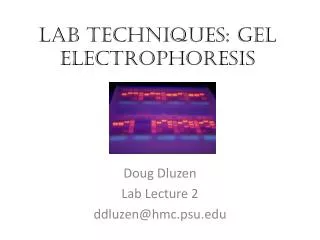
Detection techniques for gel-based proteomics
Harini Chandra Affiliations. Detection techniques for gel-based proteomics. Detection techniques using organic and fluorescent dyes for gel-based proteomics have developed significantly over the last few years allowing researchers to carry out high-throughput studies with increased sensitivity.

Detection techniques for gel-based proteomics
E N D
Presentation Transcript
Harini Chandra Affiliations Detection techniques for gel-based proteomics Detection techniques using organic and fluorescent dyes for gel-based proteomics have developed significantly over the last few years allowing researchers to carry out high-throughput studies with increased sensitivity.
Master Layout 1 Coomassie Brilliant Blue (CBB) Pro-Q-diamond Silver staining 2 Gel-based detection techniques SYPRO Ruby Red 3 Lightining fast 4 Cyanine dyes SYPRO orange 5
Definitions of the components: 1 1. Coomassie Brilliant Blue (CBB): Coomassie blue dyes (R-250 & G-250) are low cost, organic dyes that are easy to use for staining of proteins that have been separated by electrophoretic techniques. Gels are soaked in the dye solution dissolved in methanol and acetic acid, after which excess stain is then washed off with a destaining solution. The higher affinity of proteins towards the dye molecules, allows the protein bands to be selectively stained with sensitivity of 8-100 ng without significant staining of the background. These dyes are also compatible for further MS-based applications. 2. Silver staining: Electrophoresis gels are saturated with silver ions in the form of either silver nitrate or as an ammonia-silver complex after fixing the proteins in the gel. The less tightly bound metal ions are subsequently washed off and the protein-bound silver ions are reduced to metallic silver using formaldehyde under alkaline conditions in presence of sodium carbonate or citrate buffer solution. Although as little as 1 ng of protein can be detected by silver staining, the gel-to-gel reproducibility remains an issue.Compatibility of silver stains with MS is another issue, which has however, been overcome in recently introduced silver stains. 3.SYPRO Ruby Red: This is a ruthenium-based metal chelate fluorescent stain that provides a single step protein staining procedure with low background staining in polyacrylamide gels. They have been observed to be as sensitive as the silver stains (0.25-1 ng) with the liner dynamic range extending over three orders of magnitude, thereby showing better performance than CBB and silver stains. This stain can also be combined with other dyes thereby allowing multiple detections in a single gel. 4. SYPRO orange: This dye is less sensitive than SYPRO Ruby Red but is also capable of detecting proteins in SDS-PAGE gels in a rapid single step process without the requirement for any destaining procedure. As little as 4-30 ng of protein can be detected by this fluorescent dye and it is compatible for further MS-based applications. Two other similar dyes having comparable sensitivities and similar excitation and emission wavelengths are SYPRO Red and Tangerine. 2 3 4 5
Definitions of the components: 1 5. Cyanine dyes: They are water-soluble derivatives of N-hydroxy succinimide that covalently bind the e-amino groups of a protein’s lysine residues and are spectrally resolvable as they fluoresce at distinct wavelengths. The labelled protein samples can therefore be mixed and run on a single gel, thereby eliminating the problem of gel-to-gel variations, the principle employed in difference gel electrophoresis (DIGE). Cy3, Cy5 and Cy2 having sensitivities of 0.1-2 ng are most commonly used for proteomic and MS-based applications. 6. Lightning fast/Deep purple: A fluorescence-based stain obtained from the fungus Epicoccum nigrum that can be used for detecting proteins in 1-D and 2-D gel electrophoresis with sensitivity down to 100 pg protein. Stained proteins are excited by near-UV or visible light with maximum fluorescence emission occurring at around 610nm. These dyes are suited for further use with Edman or MS applications. 7. Pro-Q-Diamond: This fluorescent dye is capable of detecting modified proteins that have been phosphorylated at their serine, threonine or tyrosine residues. They are suitable for use with electrophoretic techniques or with protein microarrays and offer sensitivity down to few ng levels, depending upon the format in which they are used. This dye can also be combined with other staining procedures thereby allowing more than one detection protocol on a single gel. 2 3 4 5
Step 1: 1 Coomassie Brilliant Blue (CBB) Pro-Q-diamond Silver staining 2 Lightining fast (Deep purple total protein stain) Gel-based detection techniques SYPRO Ruby Red 3 SYPRO orange Cyanine dyes 4 Action Description of the action Audio Narration First the central heading must be shown followed by appearance of each of the arrows and the boxes. User must be allowed to click on these headings to read more about them as given in the previous two slides. Finally the titles in the blue, grey, green, yellow and red boxes must be highlighted. Each technique shown must appear one at a time as depicted. As described in previous two slides for each of the headings. 5
Step 2: 1 Forms ionic & non-covalent interactions with proteins Coomassie Brilliant Blue (CBB) Completed electrophoresis gel 2 3 Tray Mechanical rocker Coomassie blue solution 4 Action Description of the action Audio Narration The completed electrophoresis gel is placed in a tray containing the Coomassie blue staining solution, typically R-250, that has been dissolved in an aqueous solution of ethanol and acetic acid. Negatively charged Coomassie dye interacts with proteins through ionic and non-covalent interactions. This tray is then placed on a mechanical rocker that allows for uniform contact of the gel with the solution by means of gentle rocking. Show the tray on the left followed by a blue solution in it and placement of the ‘gel’ in the solution. (Please redraw chemical structure and mechanical rocker) First show the tray on the left followed by a blue solution being filled in it. Next show the ‘completed gel’ being placed in this solution. Then show the instrument on the right and the tray with gel being placed on it. This must then be shown to rock from side to side as indicated by the arrow. 5
Step 3: 1 Coomassie Brilliant Blue (CBB) Stained gel 2 Excess stain removed 3 A 2-D gel image showing final gel pattern with stained protein bands Tray Destaining solution 4 Action Description of the action Audio Narration First show the gel being removed from the blue solution in the previous slide and being placed in a colorless solution, as shown above. This must again be placed on the ‘mechanical rocker’ shown above and as the rocking takes place, the colorless solution must slowly turn blue. Show the ‘gel’ being removed from the blue solution and being placed in a transparent solution as shown above. The stained gel is then placed in a destaining solution consisting of 50% methanol & 10% acetic acidto remove any excess dye that may be bound to the gel. The gel is finally removed and viewed by means of a scanner where the blue protein bands can be easily detected. 5
Step 4: 1 Silver staining Completed electrophoresis gel 2 3 Ag+ Ag+ Ag+ Ag+ Ag+ Tray Mechanical rocker Fixing solution (acid/alcohol) – to minimize diffusion of protein bands in gel Silver stain 4 Action Description of the action Audio Narration Show the tray on the left followed by first a pale grey solution and then a pale orange solution in it. The completed gel is first placed in a fixing solution of methanol and acetic acid that fixes the protein bands in the gel and minimizes any diffusion. This must be subjected to gentle shaking for around 30 minutes after which the silver stain solution is added. The gels are rocked gently in order to allow for proper and uniform staining. First show the tray on the left followed by a pale grey solution being filled in it and the label ‘fixing solution’. Next show the ‘completed gel’ being placed in this solution. After a while, show the grey solution being removed and being filled by a pale orange solution, ‘silver stain’. When this is done, the grey circles must appear in solution. Then show the instrument on the right and the tray with gel being placed on it. This must then be shown to rock from side to side as indicated by the arrow. 5
Step 5: 1 Silver staining Stained gel OH- HCHO 2 HCHO OH- Developing solution OH- HCHO 3 Ag+ Ag+ Ag+ Ag+ Ag+ Ag+ Final gel pattern with silverstained protein bands Ag+ + HCHO + OH- Ag+ + HCOO- + H2O 4 Action Description of the action Audio Narration Show the ‘gel’ being removed from the orange solution and being placed in the pale pink solution. First show the gel being removed from the orange solution in the previous slide and then being placed in a pale pink solution containing green ovals and orange rectangles along with the grey circles, as shown above. This must again be placed on the ‘mechanical rocker’ shown in the previous slide and rocked gently. When this is taking place, the equation in the grey box must appear. Finally the image on the right must be shown. The silver stained gels are first washed to remove any excess stain and then placed in a developing solution where the silver ions get reduced to metallic silver. Formaldehyde in an alkaline solution in presence of sodium carbonate or other alkaline buffers are commonly used for this process. This gel can then be viewed as dark bands against a light background as shown in the figure on the right. 5
Step 6: 1 SYPRO Ruby Red Completed electrophoresis gel 2 3 Tray Mechanical rocker Fixing solution 50% methanol+7% acetic acid SYPRO Ruby Red stain solution 4 Action Description of the action Audio Narration As shown in animation. The completed gel is first placed in a fixing solution of methanol and acetic acid that fixes the protein bands in the gel and minimizes any diffusion. This must be subjected to gentle shaking for around 30 minutes after which the gel is soaked in fluorescent SYPRO Ruby Red stain solution. The gel is rocked gently for uniform staining with the ruthenium-based chelate dye. First show the tray on the left with a pale grey solution in it and the label ‘fixing solution’. Next show the ‘completed gel’ being placed in this solution. After a while, show the grey solution being removed and being filled by a pale red solution, ‘SYPRO Ruby red’. Then show the instrument on the right and the tray with gel being placed on it. This must then be shown to rock from side to side as indicated by the arrow. 5
Step 7: 1 SYPRO Ruby Red Gel scanner Washing solution – 10% methanol+7% acetic acid Maximum excitation wavelength – 280 nm / 450 nm 2 Emission maxima – 610 nm 3 Final SYPRO Ruby Red stained gel pattern Decreasing molecular weight Decreasing pH 4 Action Description of the action Audio Narration Show the ‘gel’ being removed from the red solution and being placed in the pale blue solution. (Please redraw all figures.) First show the gel being removed from the red solution in the previous slide and then being placed in a pale blue solution as shown above. This must again be placed on the ‘mechanical rocker’ shown in the previous slide and rocked gently. The gel must be removed and placed below the instrument shown on top right but the pattern must not be visible. Then the violet arrows indicating light must be shown to enter the instrument as indicated followed by the red beam coming out and the gel being displayed with its pattern. The gel can be left overnight in the dye solution and then washed with a methanol-acetic acid solution. The MS-compatible gel is then viewed using UV or Laser-scanning instruments with an excitation wavelength near 280 or 450 nm. The stain has two excitation maxima and an emission maxima of 610 nm. 5
Step 8: 1 Cyanine dyes Direction of migration, increasing pH Cy3 2 Sample 1 Cy5 IEF in progress IPG strip Sample 2 3 Cy2 Internal standard (Sample 1+2) 4 Action Description of the action Audio Narration As shown in animation. (Please redraw all figures.) First show the ‘sample 1’ tube followed by the red dye which must mix with it to give the red tube. Then the ‘sample 2’ tube mixing with the blue dye and then ‘sample 3’ mixing with the green dye. These 3 colored tubes must then be mixed to give the violet tube. The micropipette must be dipped in the purple solution and the yellow tip must become purple. It must then move to the blue strip and a small purple layer must appear on top of the circle on the blue strip. There must be formation of bands in the strip which must move in the direction indicated and the figure on right must be shown. In this detection technique, the dyes are mixed with the samples prior to electrophoresis. Each protein sample as well as the internal standard is labeled with a differently fluorescing cyanine dye which allows all protein samples to be simultaneously run on a single 2-DE gel. 5
Step 9: 1 Cyanine dyes DIGE Imager IPG strip Cy2 excitation wavelength 2 Cy3 excitation wavelength Cy5 excitation wavelength Direction of migration 3 All proteins with Cy2 label SDS-polyacrylamide gel slab Only Cy3 labeled proteins Only Cy5 labeled proteins 4 Action Description of the action Audio Narration (Please redraw all figures.) First show the horizontal blue strip and the blue slab below. The strip must move down and fit into the slot below. The blue bands must be shown to be moving in the direction indicated. The instrument on top right must then be shown followed by the appearance of the three beams with their respective blue figures and labels below. As shown in the animation. This gel can be viewed by illuminating it alternately with excitation wavelengths corresponding to the various Cy dyes. This technique minimizes gel-to-gel variations and allows a large number of samples to be run using fewer gels, thereby proving to be extremely useful for large-scale proteomic applications. 5
Step 10: 1 Pro-Q-diamond ProQ diamond stain 2 1. Fixing (methanol+acetic acid) 2. Staining 3. Washing (methanol+acetic acid) 3 SYPRO-Ruby Red 4 Dual staining: Pro-Q-Diamond and SYPRO Ruby Red Action Description of the action Audio Narration First show the first two trays on the left with the solution in it. The gels must be placed in these two trays. Next, show this solution being removed and the next solution as shown in middle panel being added to the tray. Finally, this solution must also be removed and the last blue solution must be added to the tray. ProQ-diamond is a useful technique for detection of phosphoproteins and when used in combination with SYPRO-Ruby red, it gives a good comparative profile of the total protein content and the phosphoprotein content. The procedure for staining is similar to SYPRO-Ruby Red as discussed earlier. This fluorescent dye offers sensitivity down to ng levels and detects phosphorylations taking place at serine, threonine and tyrosine residues. As shown in animation. 5
Step 11: 1 Pro-Q-diamond: Dual staining Gel scanner 2 Emission maxima – 580 nm Emission maxima – 610 nm 3 Phosphoprotein image Total protein image Fluorescence Fluorescence Decreasing molecular weight Phosphoprotein image Total protein image by SYPRO-Ruby Red Decreasing pH 4 Action Description of the action Audio Narration (Please redraw all figures.) First show the instrument on top followed by the yellow beam and the gel placed below that. This must be followed by the graph on the top right. Next, show the red beam coming out of the instrument with its gel below and the second graph on bottom right. As shown in the animation. The two stained gels can be scanned at their respective excitation wavelengths for detection of total protein and phosphoprotein images. The gel stained with Pro-Q-diamond will show fewer bands than the SYPRO-Ruby stained gel due to fewer number of phosphoproteins in the total protein mixture. Hence, a good comparative analysis can be done using this protocol and the stained gel can be used for further MS-based applications 5
Interactivity option 1:Step No:1 1 2 3 4 Interactivity Type Results Boundary/limits Options The rows must be displayed one at a time to the user along with its input values. User must drag and drop the inputs in the table into the correct column. Once the user drags all the inputs into their correct cells as shown above, the message ‘Well done!’ must be displayed and user is re-directed to step 2. Drag and drop. 5
Interactivity option 1:Step No:2 1 2 3 4 5
Questionnaire 1 1. Which of the following detection methods is mostlynot compatible for MS applications? Answers: a) SYPRO-Ruby red b) Pro-Q-Diamond c) Silver staining d) SYPRO-Tangerine 2. The emission maxima of Pro-Q diamond is Answers: a) 610 nm b) 580 nm c) 480 nm d) 250 nm 3. Which of the following detection stains is obtained from a natural source? Answers: a) Coomassie blue b) SYPRO-Orange c) Cyanine dyes d) Deep purple/Lightning fast 4. If a Pro-Q-labeled gel shows 8 bands and a SYPRO-Ruby red-labeled gel shows 36 bands, then how many non-phosphorylated proteins are present in the mixture? Answers: a) 44 b) 8 c) 26 d) 28 2 3 4 5
Links for further reading Reference websites: Steinberg, T. H. et al. Pro-Q Diamond phosphoprotein stain: a new reagent for detection of phosphoproteins and phosphopeptides in polyacrylamide gels and in microarrays. Research papers: • Patton, W. F. Detection technologies in proteome analysis. J. Chromatogr. B. 2002, 771, 3-31. • Mackintosh, J. A. et al. A fluorescent natural product for ultra sensitive detection of proteins in one-dimensional and two-dimensional gel electrophoresis. Proteomics 2003, 3, 2273-2288. • Johnson, C. J., Zhukovsky, N., Cass, A. E. G., Nagy, J. M. Proteomics, nanotechnology and molecular diagnostics. Proteomics 2008, 8, 715-730. • Berggren, K. N. et al. An improved formulation of SYPRO Ruby protein gel stain: Comparison with the original formulation and with a ruthenium II tris (bathophenanthroline disulfonate) formulation. Proteomics 2002, 2, 486-498. • Chakravarti, B., Ratanaprayul, W., Dalal, N., Chakravarti, D. N. Comparison of SYPRO Ruby and Deep Purple Using Commonly Available UV Transilluminator: Wide-Scale Application in Proteomic Research. J Proteome Res. 2008, 7(7), 2797-802.