Docking molecules with Vina
Docking molecules with Vina. Autodock Vina. To study molecules they must be docked. Docked molecules bind their enzyme or receptor in a specific conformation Docked molecules bind with high affinity The best way to get docked conformation is to use X-ray crystallography

Docking molecules with Vina
E N D
Presentation Transcript
To study molecules they must be docked • Docked molecules bind their enzyme or receptor in a specific conformation • Docked molecules bind with high affinity • The best way to get docked conformation is to use X-ray crystallography • Alternative: use docking software like Vina to predict docked conformation
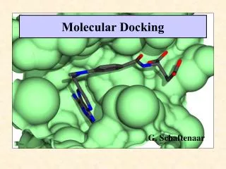
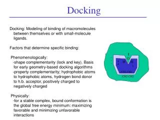
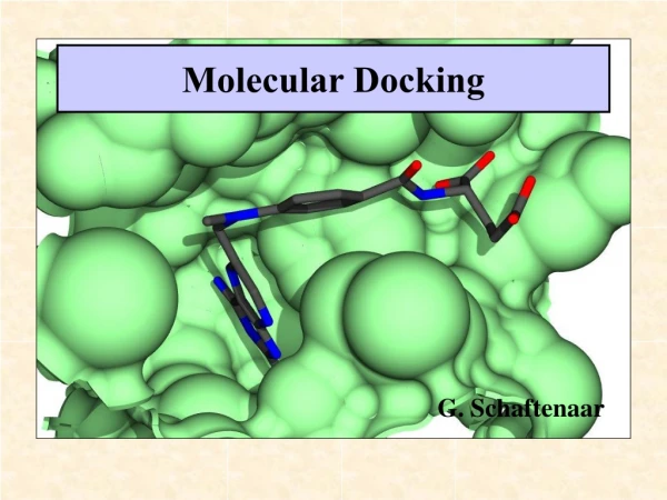
What is docking? • Find the conformation with which a ligand binds a receptor • This information includes full coordinates of all ligand atoms • The docked configuration is the (presumed) lowest energy binding site
Steps • 1. get protein structure • 2. create files with protein only • 3. Use a known ligand (not your ligand) to define the ligand binding site • 4. Generate some modified structure files • 5. Use files with docking software • 6. Extract predicted conformation and make new file with protein and target ligand docked
Make a folder • Need a place to store many files • Make c:\thesis
Get PDB file • Go to PDB RSCB on web • Search for PDB code if unknown • Once PDB code is known, download it • Use PDB/Tools, download entries and follow instructions • You must have Java enabled • Change mmCIF to PDB as type of file • Change compressed to uncompressed • Save in your thesis folder
Start with PDB file with protein and ligand (not your ligand) • Make a copy of the PDB file • Split it into only protein and only ligand • Ligand will be used to get coordinates of binding site • Example: beta adrenergic receptor and carazolol • 2rh1.pdb
Use wordpad text editor • Copy cau (= carazolol, ligand) from 2rh1 • Paste into a file called car.pdb • REMARK carazolol • HETATM 3598 O17 CAU A 408 -33.477 10.957 8.170 1.00 50.96 O • HETATM 3599 C16 CAU A 408 -32.267 10.230 8.041 1.00 45.65 C • HETATM 3600 C18 CAU A 408 -32.478 8.951 7.225 1.00 51.24 C • HETATM 3601 N19 CAU A 408 -33.702 8.250 7.600 1.00 54.99 N • HETATM 3602 C20 CAU A 408 -33.806 6.805 7.498 1.00 60.13 C
PDB files have 3D structure information including XYZ coordinates of atoms • Also have atom numbers • Residue numbers • Atom types
Find the center of the ligand • This will be the center used when you dock your ligand to the receptor • Start with the X coordinate. Add all the X values and then divide by the number of atoms. Record for later • Repeat for Y and Z coordinates • The only purpose of having a ligand at this point is to define the ligand binding site.
Find coordinates • z • y • x • REMARK carazolol • HETATM 3598 O17 CAU A 408 -33.477 10.957 8.170 1.00 50.96 O • HETATM 3599 C16 CAU A 408 -32.267 10.230 8.041 1.00 45.65 C • HETATM 3600 C18 CAU A 408 -32.478 8.951 7.225 1.00 51.24 C • HETATM 3601 N19 CAU A 408 -33.702 8.250 7.600 1.00 54.99 N • HETATM 3602 C20 CAU A 408 -33.806 6.805 7.498 1.00 60.13 C
Visualizing structures • Try rasmol or raswin • Download at google: rasmol bernstein • Several web tutorials available
Adding hydrogens • Hydrogen is rarely found in PDB files • But all atoms are needed to dock molecules • Solution: add back missing hydrogens • We will use Babel
OpenBabel • Download openbabel from its web site • Install • Use the command prompt to run the program • Use a full ‘path’, that is a description of the program location • Add hydrogens • On my machine that is (one line): • \lm\downloads\openbabel-2.1.1\babel.exe -ipdb car.pdb –opdb carH.pdb -h
Check that hydrogens have been added • HETATM 20 C1 CAU 408 -26.395 6.432 7.689 1.00 0.00 C • HETATM 21 C6 CAU 408 -27.717 6.731 8.006 1.00 0.00 C • HETATM 22 C5 CAU 408 -28.269 7.948 7.652 1.00 0.00 C • ATOM 23 H CAU 408 -34.319 10.255 8.260 1.00 0.00 H • ATOM 24 H CAU 408 -31.904 9.929 9.079 1.00 0.00 H • ATOM 25 H CAU 408 -31.591 8.256 7.396 1.00 0.00 H
Other ligands • Up to now we have been dealing with the ligand found in the PDB file with your receptor/protein • You may wish to try docking that ligand as a control • At some point you will want to deal with other ligands
Getting other ligands • To use ligands they must be transferred from the page to PDB format • Some ligands may be present in other files in the protein data bank – try searching • Ligands can be drawn, e.g. with DS Viewer Pro or other software and saved in PDB format • Most ligand files will have to have hydrogens added
Add hydrogen to the protein • Repeat ligand procedure, but with protein • We did car.pdb carH.pdb • Now make copy of the protein pdb file with the ligand deleted (e.g. CAU in this example) • (you can delete WAT and other ligands too) • Now, with full path, use babel to add H
Add hydrogen to protein • Full path on my machine (yours will differ) • \lm\downloads\babel-2.1.1\babel.exe –ipdb bar.pdb –opdb barH.pdb -h • Result: bar.pdb barH.pdb
We’re almost ready to dock • But we need two things: • Torsions • Hydrogen bonds
Torsions • The ligand may have rotatable bonds • Benzene can not twist (0 torsions) • Hexane can twist (3 torsions, we’ll ignore rotating methyl groups) • MGLtools calculates these • Vina will twist the ligand to try to find the best fit during docking
H bonds • We need to find H bond acceptors and donors • MGLtools also calculates these
MGLtools • MGLtool, ADT, Autodock tools are the same • Download from MGLtools site • Install • Read tutorial • Click to open window
MGLtools We will use MGLtools/Autodock tools for two things: Annotate the ligand(s) Annotate the receptor
Autodock / MGL tools • First we will process the ligand to add torsions and H bond information • On left side of ADT screen find ligand menu • Open ligandH.pdb (or other file) • Note: this is the ligand file you want to dock, not the X-yl structure file, unless you are docking it as a control
Autodock/MGLtools and ligand • Select torsion tree • Find the ligand torsion ‘root’. Detect root • Aromatic carbons: set names • Output: Save file as carH.pdbqt • It is key that you save the pdbqt file that MGLtools makes
ADT and protein/receptor • Find ‘grid’ menu • Macromolecule • Open receptor file with hydrogens (receptorH.pdb) • The file gets processed • Save as ligandH.pdbqt • Do not choose ‘flexible’
ADT receptor refinement • Now the receptor file and ligand file have been saved as .pdbqt files. • These have H bond and torsion information • Look at the ligandH.pdbqt file using wordpad or simpletext • It still looks like a .pdb file, but has extra information added
Now we have all of the files that we need to dock a ligand • Let’s check: • receptorH.pdbqt • ligandH.pdbqt • Binding site coordinates
Setting up Vina • Vina needs to know what you want to do: • What receptor • What ligand • Where the binding site is • Where to send out the results • This information is placed in a file called config.txt
Configuration file, config.txt • Config.txt gives information to Vina as it docks your ligand • The left side of each = is Vina code (don’t change) • The right side of each = is your input • You control the site of binding, the size of the site, and the receptor and ligand
Example Vina configuration file out = out_carh.pdbqt receptor = 2rhe.pdbqt ligand = carh.pdbqt center_x = -28 center_y = 9 center_z = 6 size_x = 25 size_y = 25 size_z = 25 energy_range = 4
Download Vina • Web site AutoDock Vina • Install in ‘thesis’ folder, or wherever your config file and .pdbqt files are saved. • Get the right version of Vina for your computer (e.g. PC vs. Mac)
Dock ligand • Open command prompt • Make certain that command prompt is pointed to thesis directory/folder • Use the cd command with command prompt or Mac command screen
Finding the right directory • If necessary change directory using ‘cd’ • E.g. type ‘cd \thesis’ [without the quotes] for a PC • Or type ‘cd /thesis’ [without quotes] for mac • This assumes that you put all your files in the directory ‘thesis’ on the c:drive or equivalent • Type dir/p [PC] or ls [Mac] to check that your files are present
Vina docking To dock, type : vina.exe --config config.txt
Vina output • It takes a minute or two for Vina to dock • This represents millions of docked positions being analyzed • The output file name is defined by the config file out = outfilename.txt • Each outfile has several models • We usually only care about model 1, the best
Output score • We often want to know how tightly a ligand binds • Vina gives an estimate in the output file • Look for Vina Result: x • If x is -10 or less, binding is very tight • If x is -6 to -7 binding is just random, not tight • In between is hard to judge