Max F. Perutz Laboratories
460 likes | 709 Vues
Max F. Perutz Laboratories. Georg Weitzer. Kapitel 2 Über die in vitro Differenzierung von embryonalen Stammzellen: Embryoid Bodies – ein Modell für die frühe Embryogenese?. Medical University of Vienna. Georg Weitzer. Wie entstehen somatische Zellen in Embryoid Bodies?

Max F. Perutz Laboratories
E N D
Presentation Transcript
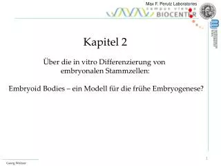
Max F. Perutz Laboratories Georg Weitzer Kapitel 2 Über die in vitro Differenzierung von embryonalen Stammzellen: Embryoid Bodies – ein Modell für die frühe Embryogenese?
Medical University of Vienna Georg Weitzer • Wie entstehen somatische Zellen in Embryoid Bodies? • Ist die Gastrulation chaotisch, oder gibt es reproduzierbare morphologische Strukturen?
Medical University of Vienna Georg Weitzer Entwicklungsabschnitte in vivo und in vitro • Pre-implantations Entwicklung: Tag 0 - 4 • Pre-gastrulations Entwicklung : Tag 4 - 7 • Gastrulation / Keimblattbildung: Tag 7 - 9… In der Maus
Medical University of Vienna Georg Weitzer 20 min. Trypsin 24 Stunden Herstellung von Embryoid Bodies
Trophectoderm Inner Cell Mass Medical University of Vienna Blastocyst 6-7 cell divisions Primitive Endoderm Primitive Ectoderm (Epiblast) (Hypoblast) Day 2 -3 Day 4 Day 1 Day 1.5 Embryonic Stem Cells Embryoid Bodies Georg Weitzer • Pre-implantation development: Day 0 – 4 Morphological evidence In vivo Zygote In vitro
Medical University of Vienna Georg Weitzer Embryoid Bodies Tag 1-3 Tag 4,5 Tag 5.5 Anna Wobus, Gatersleben, D Erfinderin der von ESC abstammenden Embryoid Bodies Tag 6.5
Medical University of Vienna Georg Weitzer Der Hypoblast und Epiblast bildet sich, aber kein Trophektoderm. Embryoid Bodies verhalten sich wie die Innere Zellmasse. Kompaktierung der ESCs kann nicht wirklich mit der Kompaktierung der Blastomere verglichen werden.
Medical University of Vienna Georg Weitzer • Pre-implantation development: Day 0 - 4 Molecular evidence Genexpression in Embryiod Bodies typisch für: Primitives Endoderm Primitives Ektoderm
Implantation Pseudo-Implantation Medical University of Vienna Blastocyst In vitro Primitive Endoderm Visceral Endoderm Parietal Endoderm Embryoid Bodies Georg Weitzer 2. Pre-gastrulation development: Day 4-7 Morphological evidence In vivo =
Medical University of Vienna Extra-embryonales Gewebe bildet sich genau so, wie um den Embryo. Georg Weitzer 2. Pre-gastrulation development: Day 4-7 Morphological evidence In vitro Visceral Endoderm Dolichos biflorus agglutinin Pseudo-implantation Parietal Endoderm ß-catenin
Egg cylinder stage Medical University of Vienna Prim. Ectoderm Primitive Mesoderm Cardiocytes Day 8 Embryoid Body Erythrocytes Day 6 Georg Weitzer In vivo 3. Gastrulation / Germlayer formation: Day 7-9... Morphological evidence Primitive streak stage Head fold stage In vitro
Medical University of Vienna Georg Weitzer Der zeitliche Ablauf der Keimblattentwicklung in vitro ist gleich wie bei der Gastrulation in vivo.
Cardiomyocytes 3. Gastrulation / Germlayer formation: Day 7-9… Molecular evidence Genexpression in Embryiod Bodies typisch für: Medical University of Vienna Definitive Endoderm Primitive Mesoderm Definitive Georg Weitzer
Parietal Endoderm Visceral Endoderm
Mesoderm Primitve Ectoderm
Medical University of Vienna Georg Weitzer = Ist die Entstehung der somatischen Zellen während der Gastrulation chaotisch?
Point symmetry Line symmetry Medical University of Vienna Georg Weitzer Development of “implanted” embryoid bodies Day 6 Day 6.5 Day 7.0 Day 8.0 Day 9.0
Medical University of Vienna ? Georg Weitzer Mesodermbildung in Embryoid Bodies Primitve Mesoderm Brachyury Cardiomyocytes MHCa
Medical University of Vienna In 65 +/- 7 % der Embryoid Bodies beginnen die ersten Kardiomyozyten „links unten“ zu schlagen! (N= 349) Embryoid Bodies sind asymetrisch! Georg Weitzer left right Braking line symmetry Area where mesodermal cells emerge upper lower
Medical University of Vienna Georg Weitzer
Max F. Perutz Laboratories Max F. Perutz Laboratories Georg Weitzer Nachtrag zur Entwicklung von Embryoid Bodies Figure 3. Xu Scientific Report 2004 Schematic illustration of GATA factors in the perception of differentiation cues. Embryonic stem cells can be induced in vitro to differentiate by either aggregation to form embryoid bodies or by retinoic acid. Upon aggregation, embryonic stem cells differentiate into primitive endoderm, an epithelial cell type, covering the surface of the spheroids. Disabled-2 (brown staining/arrow) is a marker for primitive endoderm cells.
Nachtrag zur Entwicklung von Embryoid Bodies Extra-cellular matrix Provides indispensable signals for epiblast formation in vitro. Fig 1. David Edgar, Liverpool Without a basement membrane (laminin -/-), the concentric ectodermal and endodermal epithelia fail to develop in ES cell-derived embryoid bodies (A, C). On rescue of BM (B, D), the epithelia form and cell death creates a pro-amniotic cavity at the centre of the embryoid body. C and D are confocal images showing cells (blue) and BM (green).
Entwicklung von Embryoid Bodies Figure 8. Disorganized ectoderm in cystic EBs derived from afadin-/- ES cells. Cystic EBs were subjected to histological analysis or immunofluorescence microscopy. For immunofluorescence microscopy, cystic EBs were doubly stained with rhodamine-phalloidin and the polyclonal anti–l-afadin antibody, or with the anti–E-cadherin and anti–ZO-1 antibodies. (A) Cystic EBs derived from wild-type ES cells; (B) cystic EBs derived from afadin-/- ES cells; (Aa and Ba) microscopic appearance; (Ab and Bb) staining with hematoxylin and eosin; (Ac, Ad, Bc, and Bd) double staining with rhodamine-phalloidin and the anti–l-afadin antibody; (Ac and Bc) l-afadin; (Ad and Bd) F-actin; (Ae, Af, Be, and Bf) double staining with the anti–E-cadherin and anti–ZO-1 antibodies; (Ae and Be) E-cadherin; and (Af and Bf) ZO-1. Arrowheads indicate junctional complex regions. en, endodermal layer; cy, cyst cavity; ec, ectodermal layer; rm, Reichert's membranes; and cm, cell mass. Bars, 150 µm (Aa and Ba); 100 µm (Ab and Bb); and 30 µm (Ac–Af and Bc–Bf
Embryoid bodies erlauben eine relative einfache Bestimmung der Potentialität von Stammzellen - und über dies hinaus - die Untersuchung von molekularen und zellulären Prozessen während der Embryogenese, die experimentell im Embryo nicht erfassbar sind.
Murine Embryo, day 7 Embryoid Body, day 7 Medical University of Vienna Embryonic Stem Cells Blastocyst, day 3.5 ? In vivo In vitro Georg Weitzer
Eine Kolonie von CardiovascularProgenitor Cells
Figure 5 | Transcription factor cross-antagonisms in a cascading landscape of unstable and stable cell states. The territory, represented as a mountain range, depicts all possible solutions of a single regulatory network that specifies cell identity. Robust network states correspond to stably differentiated cell types (deep basins in the low-lying plains) whereas unstable solutions correspond to ridges and slopes in the landscape. The latter are only fleetingly occupied during development and thus unlikely to correspond to observable cell types. ES cells, embryonic stem cells; HSCs, haematopoietic stem cells.
Figure 5 | Transcription factor cross-antagonisms in a cascading landscape of unstable and stable cell states. The territory, represented as a mountain range, depicts all possible solutions of a single regulatory network that specifies cell identity. Robust network states correspond to stably differentiated cell types (deep basins in the low-lying plains) whereas unstable solutions correspond to ridges and slopes in the landscape. The latter are only fleetingly occupied during development and thus unlikely to correspond to observable cell types. ES cells, embryonic stem cells; HSCs, haematopoietic stem cells.
Zellfusion und Plastizität Donor bone marrow cells with a ß-actin-promoter—loxP—STOP—loxP—LacZ transgene ß-actin-Promoter::CRE mice as recipients If cell fusion takes place, tertraploid cells have both CRE and the transgene Cre generates a ß-actin-promoter—loxP—LacZ transgene Fused cells become blue. Bond et al., 2009, siehe Lernunterlagen