Understanding Protein Targeting and Vesicle Budding: Insights into Molecular Mechanisms
This article explores the molecular basis of protein targeting, particularly in relation to bipolar disorder. It discusses how proteins synthesized in the cytosol are sorted and delivered to membranes and organelle lumens, specifically focusing on the mechanisms of Sec and SRP-mediated translocation into the ER. Key topics include protein budding, the role of clathrin, COPI, COPII, and the SNARE hypothesis, as well as endocytosis processes like phagocytosis and pinocytosis. Understanding these processes is crucial for elucidating the molecular underpinnings of cellular functions and potential dysfunctions.

Understanding Protein Targeting and Vesicle Budding: Insights into Molecular Mechanisms
E N D
Presentation Transcript
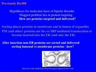
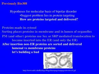
Previously Bio308 • Hypotheses for molecular basis of bipolar disorder • Suggest problem lies in protein targeting • How are proteins targeted and delivered? Proteins made in cytosol Sorting places proteins in membrane and in lumen of organelles PM (and other) proteins use Sec or SRP mediated translocation to become inserted into the ER (and only the ER) After insertion non-ER proteins are sorted and delivered lumenal vs membrane proteins let’s building a bud http://www.udel.edu/Biology/Wags/histopage/empage/ebv/ebv10.gif
Budding CBI 13.1 Clathrin Fig 17-58
Coat Components Clathrin COPI COPII Identity determined by what the vesicle contains and it’s coat. http://userpage.chemie.fu-berlin.de/biochemie/aghaucke/clath.jpg
Highly hydrophobic + charged - charged Hydroxylated Other Sar1p-GTP form exposes helix that anchors protein to ER surface by ‘floating’ with hydrophobic a.a. interacting with membrane core Budding II ER vesicle budding Sar1p N-terminal helix Amino Acid Key Drin, G, and B. Antonny (2005) News and Views: Helices sculpt membrane. Nature vol: 437
Budding III ER vesicle budding Floating many Sar1p in top leaflet makes it ‘bigger’ than the bottom one. Results --> bulge that can more easily interact with coat proteins. Drin, G, and B. Antonny (2005) News and Views: Helices sculpt membrane. Nature vol: 437
ER vesicle budding….fission Fission Ring of parallel helices at neck might aid fission. New data for ER; had seen a protein (epsin) help deform PM for clathrin coated vesicles. May suggest that using a helix to deform membrane is common mechanism for budding/fission
Targeting/Docking: What happens after budding? How do vesicles dock with specific target membrane? http://dir2.nichd.nih.gov/nichd/cbmb/sob/in_vivo_dyn.html
The SNARE hypothesis V-SNARE T-SNARE Role of p115 Role of Rab proteins retrograde Fig 17-59
Synaptic vesicle fusion VAMP Syntaxin SNAP 25 Rab3a Synaptotagmin
Moving in the other direction: endocytosis Types: Phagocytosis– specialized cells Pinocytosis– all cells Connection– perhaps the # of our receptor’s on PM is controlled by endocytosis Pinocytosis ‘problem’ rate of pinocytosis internalizes 100% of PM per hour ? (How can this be?)