The Shoulder Complex
The Shoulder Complex. Chapter 22 Westfield High School Houston, Texas. Anatomy. Clavicle – Collar Bone Scapula – Shoulder Blade Humerus. Articulations. Sternoclavicular – SC joint. Sternum and Clavicle. Acromioclavicular – AC joint, Acromion process of the scapula and the clavicle

The Shoulder Complex
E N D
Presentation Transcript
The Shoulder Complex Chapter 22 Westfield High School Houston, Texas
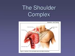
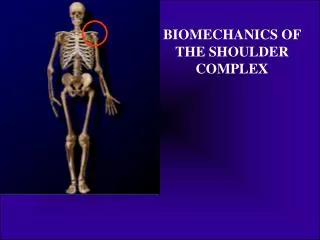
Anatomy • Clavicle – Collar Bone • Scapula – Shoulder Blade • Humerus
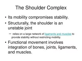
Articulations • Sternoclavicular – SC joint. Sternum and Clavicle. • Acromioclavicular – AC joint, Acromion process of the scapula and the clavicle • Glenohumeral Joint – ball and socket joint, large ball and small socket, like golf ball on a golf tee, more mobility means less stability
Articulations • Scapulothoracic Joint – not a true joint, movement of the scapula over the thoracic wall.
Prevention of Shoulder Injuries • Proper physical conditioning • Full ROM should be used with conditioning • Proper warm up and stretching • Instruction on falling properly, not on outstretched arm, but a shoulder roll • Properly fitting shoulder pads in collision sports • Correct techniques for sports with overuse injuries • Throwing, spiking, overhead smashing, tackling, blocking swimming
Injuries to Shoulder Complex • Clavicle Fractures: one of the most frequent fractures in sport, result from fall on an outstretched arm or a direct impact. • The majority of clavicle fractures occur in the middle third from a direct impact. In young athletes these fractures are usually of the greenstick type.
Injuries to Shoulder Complex • Clavicle Fracture: The athlete will usually support the arm on the injured side and tilt his head toward that side, with the chin turned to the opposite side. During inspection the injured clavicle appears slightly lower then the unaffected side. Deformity may also be felt during palpation.
Injuries to Shoulder Complex • Clavicle Fracture: Treatment • X-Rays • Physician performed reduction • Immobilization with figure eight wrap • Immobilization for 6 to 8 weeks • Some may need surgical repair
Injuries to Shoulder Complex • Fractures to Humerus: • Can happen to the shaft, proximal humerus, and the head of the humerus • How the fracture occurs differs for each type of fracture
Fractures to Humerus • Humeral Shaft: usually caused by a direct blow or a fall on the arm. Most mid –shaft fractures are comminuted or transverse fractures and a deformity is often produced because the bone fragments override each other as a result of sting muscular pull.
Fractures to Humerus • Mid-Shaft Fractures: the raidal nerve can be severed from fragments. If so, the athlete will show wrist drop and can not supinate forearm. • X-Ray and treat by physician to eliminate possible nerve damage.
Fractures to Humerus • Proximal Fracture: pose considerable danger to nerves and vessels of that area. Can result from a direct blow, a dislocation, or the impact received by falling onto the outstretched arm. This fracture may be mistaken for a shoulder dislocation. The greatest number of fractures take place at the surgical neck.
Fractures to Humerus • Epiphyseal Fracture: a fracture to the head of the humerus. More common in the young athlete. Usually ten years of age or younger. Caused by direct blow or by an indirect force traveling along the length of the axis of the humerus.
Fractures to Humerus • Management: Difficult to see a fracture to the humerus, so get X-Rays • Signs include: • Pain • Inability to move arm • Point tenderness • Discoloration • Possibility of paralysis
Fractures to Humerus • Treatment: • Removal from competition • Referral to physician • Immediate support • Immobilization • Can heal in 2 to 6 months
Sternoclavicular Sprain • Are relatively uncommon, but can occur from mainly falling on the shoulder or a direct blow to the SC joint. • Grade I: little pain and disability, with some point tenderness but no deformity. • Grade II: displays subluxation of the SC joint with visible deformity, pain, swelling, point tenderness and inability to abduct the shoulder in full ROM or to bring the arm across the chest.
Sternoclavicular Sprain • Grade III: most severe, with complete dislocation with gross displacement of the clavicle at its sternal junction, swelling, and disability, indicating complete rupture of the sternoclavicular and costoclavicular ligaments. If displaced posteriorly, pressure may be placed on the blood vessels, esophagus, or trachea, causing a life-or-death situation.
Sternoclavicular Sprain • MANAGEMENT: • RICE • Physician visit to reduce any displacement • Immobilization for 3 to 5 weeks • There is a high incidence of reoccurrence of SC sprains
Acromioclavicular Sprain • Extremely vulnerable joint • Most often induced by direct force to the acromion process that forces it downward, backward, and inward while the clavicle is pushed down against the rib cage. • May also be injured when an upward force is exerted against the long axis of the humerus by a fall on an outstretched arm.
Acromioclavicular Sprain • Prevention includes proper fitting of protective equipment, conditioning to provide a balance of strength and flexibility to the entire shoulder complex, and teaching proper falling techniques. • A contusion to the distal of the clavicle can be mistaken for Grade I AC sprains.
Acromioclavicular Sprain • Grade I: point tenderness and discomfort during movement; no disruption of the A/C joint, indicating only mild stretching of the A/C and coracoclavicular ligaments.
Acromioclavicular Sprain • Grade II: tearing or rupture of the A/C ligaments with associated stretching of the coracoclavicular ligament; there is a partial displacement and prominence of the lateral end of the clavicle when compared to the uninjured side; point tenderness during palpation of the injury site and the athlete is unable to to abduct through full ROM or to bring the arm completely across the chest.
Acromioclavicular Sprain • Grade III: involves complete rupture of the acromioclavicular and coracoclavicular ligaments. • Grade IV: exhibits posterior dislocation of the clavicle with complete disruption of the acromioclavicular ligament. Some Grade IV sprains will have the coracoclavicular ligament intact.
Acromioclavicular Sprain • Grade V: there is complete loss of both acromioclavicular and coracoclavicular ligaments in addition to tearing of the trapezius and deltoid attachment to the clavicle and acromion. Gross deformity and prominence of the distal clavicle, severe pain, loss of movement, and instability of the shoulder complex.
Acromioclavicular Sprain • Grade VI: vary rare in the athletic setting and involves the clavicle being displaced inferior to the coracoid behind the coracobrachialis tendon.
Acromioclavicular Sprain • Management: three basic procedures: • Application of cold and pressure to control local hemorrhage • Stabilization of the joint by a sling and swathe bandage • Referral to physician to definitive diagnosis and treatment
Acromioclavicular Sprain • Management: • Grade I: use sling for three or four days • Grade II: 10 to 14 days of protection in a sling • Grade III: non-operative with approx. 2 weeks of protection in a sling • Grade IV through VI: require surgical intervention using open reduction with internal fixation
Shoulder Dislocations • An anterior glenohumeral dislocation may result from direct impact to the posterior or posteriorlateral aspect of the shoulder. The most common mechanism is forced abduction, external rotation, and extension that forces the humeral head out of the glenoid cavity.
Shoulder Dislocations • Posterior glenohumeral dislocation is usually forced adduction and internal rotation of the shoulder or a fall on an extended and internally rotated arm.
Shoulder Dislocations • Anterior dislocation displays a flattened deltoid contour. Athlete will carry the arm in slight abduction and external rotation and is unable to touch the opposite shoulder with the hand of the affected arm. There is moderate pain and disability.
Shoulder Dislocations • Posterior dislocations will produce severe pain and disability. The arm is often held in adduction and internal rotation. The anterior deltoid muscle is flattened, the acromion and coracoid processes are prominent, and the head of the humerus also may be seen posteriorly. There is limited external rotation and elevation.
Shoulder Dislocations • MANAGEMENT: • Immediate immobilization in a comfortable position using sling with a folded towel or small pillow placed under the arm • Immediate reduction by a physician • Ice to control hemorrhage • X-Rays before reduction • Reconditioning
Shoulder Dislocations • Treatment: • Strengthen all muscles around the shoulder joint • Internal rotation • External Rotation • Long head of the biceps • Empty the can • Wear harness for playing athletics