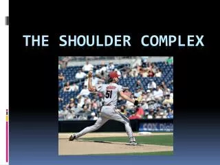
SHOULDER COMPLEX
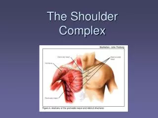
SHOULDER COMPLEX. Extrinsic Shoulder Muscles. Review the following: Pectoralis major Pectoralis minor Serratus anterior Trapezius complex Latissimus dorsi Levator scapulae Rhomboideus major Rhomboideus minor. Sensory Supply to Shoulder. Supraclavicular nerves:

SHOULDER COMPLEX
E N D
Presentation Transcript
Extrinsic Shoulder Muscles • Review the following: Pectoralis major Pectoralis minor Serratus anterior Trapezius complex Latissimus dorsi Levator scapulae Rhomboideus major Rhomboideus minor
Sensory Supply to Shoulder • Supraclavicular nerves: From cervical plexus C3-4 Supply skin over clavicle and over the superior-lateral aspect of pectoralis major • Cutaneous branches of dorsal rami: Penetrate deep and superficial back muscles Supply skin on either side of midline of back
Shoulder Movements • Elevation (scapula): Levator scapulae and rhomboids • Depression (scapula): Latissimus dorsi
Shoulder Movements • Abduction (scapula): Serratus anterior • Abduction (shoulder): Middle deltoid Biceps brachii assists • Adduction (scapula): Middle trapezius • Adduction (shoulder): Pectoralis major (clav) Latissimus dorsi
Shoulder Movements • Upward rotation (scapula): Serratus anterior Upper and lower trapezius (force couple) • Downward rotation (scapula): Rhomboids Levator scapulae
Quadrilateral Space • Boundaries: Inferior glenohumeral capsule. Teres major. Triceps longus. Surgical neck of humerus. • Contents: Axillary nerve. Posterior circumflex humeral artery.
Shoulder Complex Joints • Sternoclavicular joint (SC) • Acromioclavicular joint (AC) • Glenohumeral joint (GH)
Sternoclavicular Joint (SC) • Plane synovial joint • Articulating surfaces: Sternal end of clavicle Articular notch on manubrium First costal cartilage • 3 degrees of freedom
SC Joint Movements • Elevation and depression: Occurs around AP axis Elevation = 45 degrees Depression = 15 degrees • Protraction and retraction: Occurs around vertical axis Protraction ROM = 15 degrees Retraction ROM = 15 degrees • Rotation: Occurs around transverse axis
Sternoclavicular Joint • Joint disc Fibrocartilage meniscus Attached to: Clavicle inferiorly Manubrium and first costal cartilage inferiorly • Ligaments: Anterior/posterior sternoclavicular ligaments: Check anterior/posterior movements of clavicular head.
SC Joint Ligaments • Costoclavicular ligament: Axis for elevation and depression Axis for protraction and retraction Main check for elevation • Interclavicular
Acromioclavicular Joint • Plane synovial joint • 3 degrees of freedom • Articulation surfaces: Convex facet on lateral end of clavicle Concave facet on acromion
AC Joint Movements • Scapular rotation: Occurs around AP axis. • Winging of vertebral border of scapula: Occurs around vertical axis. • Tipping of inferior angle of scapula: Occurs around coronal axis.
Acromioclavicular Joint • Acromioclavicular ligaments: Superior and inferior Reinforce joint capsule • Coracoclavicular: Trapezoid (lateral) Conoid (medial)
Glenohumeral Joint • Ball-and-socket joint • Synovial • Components: Head of humerus Glenoid fossa of scapula • Glenoid labrum Fibrocartilage meniscus Deepens articulating surface of glenoid fossa
Glenohumeral Joint • Ligaments: Glenohumeral Coracohumeral Coracoacromial • Joint capsule: Very lax Up to an inch of passive distraction
GH Joint Movements • Flexion/extension of brachium: • Abduction/adduction of brachium: • Lateral/medial rotation of brachium:
Shoulder Complex Components • Scapulothoracic joint: Not a true anatomic joint Represented by sliding of scapula on thoracic cage • Coracoacromial arch: Components: Acromion Coracoid process Coracoacromial ligament • Subacromial/subdeltoid bursae
Rotator Cuff • Composed of four muscles whose tendons of insertion form a partial “cuff” around the head of the humerus. • Involved in snubbing and rotating head of humerus
Rotator Cuff Components • Supraspinatus S • Infraspinatus I • Teres minor T • Subscapularis S
Scapulohumeral Rhythm • Refers to relatively uninterrupted movement of upper extremity from dependent position to full abduction. • Requires simultaneous and coordinated movements of all the previous-named joints.
Scapulohumeral Rhythm • ROM: Full abduction: 180 degrees Contributed by glenohumeral joint: 120 degrees Contributed by scapulothoracic movement: 60 degrees: Sternoclavicular joint = 40 degrees Acromioclavicular joint = 20 degrees Ratio of GH to ST = 2:1
Steps in Arm Abduction • Movement (searching) of scapula: Serratus anterior clamps scapula to thoracic wall • Snubbing of head of humerus into glenoid fossa: Rotator cuff muscles • First few degrees of abduction: Supraspinatus • External rotation of humerus: Infraspinatus
Clinical Applications • Deltoid paralysis: Axillary nerve • Serratus anterior paralysis: Long thoracic nerve • Tears in rotator cuff Supraspinatus most often torn: