Download
1 / 35
420 likes | 731 Vues
Somatosensory Systems. Kimberle M. Jacobs, PhD Sanger 11-046 827-2135 kmjacobs@vcu.edu. Somatosensory Systems. Somatosensory Systems – All senses other than Olfaction, Gustation, Audition, Vision, and Vestibular Sensation: Modalities

E N D
Somatosensory Systems Kimberle M. Jacobs, PhD Sanger 11-046 827-2135 kmjacobs@vcu.edu
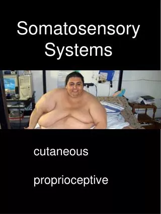
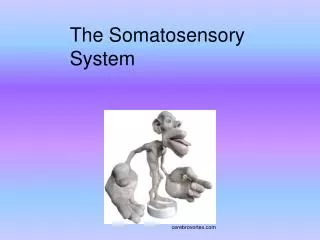
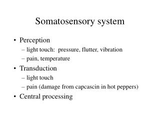
Somatosensory Systems Somatosensory Systems – All senses other than Olfaction, Gustation, Audition, Vision, and Vestibular Sensation: Modalities Touch (and Pressure), Vibration Sense, Proprioception, Kinesthesia, Stereognosis, Pain & Temperature Proprioception - sense of static and dynamic position of limbs and body Kinesthesia - the ability to feel movements of the limbs and body Stereognosis – ability to recognize objects based on touch alone • Fine Discrimination Touch • Pain and Temperature
Somatosensory Systems LEARNING NEURONAL PATHWAYS REQUIRES REPETITION • Details of pathway: Where are cell bodies located, where is the synapse made, where do axons cross the midline, first structure, second structure, etc. • Location of nuclei and tracts within brain sections
Conscious Somatosensation Spinocerebellar Tracts (IPSILATERAL) BODY HEAD Trigeminal System PAIN & Temp Fine Touch Pain Touch Lateral Spino-Thalamic Dorsal Column System Spinal Principal (Main) Non-conscious Proprioception Information reaching consciousness goes to the CONTRALATERAL Neocortex, Nonconscious Sensory Information goes to the IPSILATERAL Cerebellum
The Basic Plan for Somatosensory Information to Consciousness o Quaternary (4 ) Action Potential Initiation Site 4 Receptors can be mechanoreceptors (sensing touch), free nerve endings (sensing pain & temperature), or proprioceptors (muscle spindles & golgi tendon organs) • Adequate Stimulus – The stimulus modality to which a sense organ responds optimally. • Generator Potentials are depolarizations in receptors that are graded relative to the intensity and form of the stimulus. 3 Crossing occurs either in spinal cord OR brainstem 2 1 2nd order neuron is located either in spinal cord OR brainstem Outside the CNS!
Key Elements of the Basic Plan: SS to Consciousness o Quaternary (4 ) Action Potential Initiation Site • The basic plan for somatosensory information to consciousness involves 4 neurons and 3 synapses. • Cell bodies of the primary afferents are located outside of the central nervous system (in the dorsal root ganglion for body information and in the Trigeminal ganglion for head information). • Cell bodies of the second order neurons are located either in the spinal cord (anterolateral system) or in the brainstem (dorsal column system). • The axon of the second order neuron crosses the midline. • Cell bodies of the third order neurons are located in the thalamus (within VPL for body information and VPM for head information). • The fourth order neuron is located within the Primary Somatosensory cortex (Brodman’s areas 3, 1, 2). 4 3 2 1 Outside the CNS!
Before = IPSI, After = CONTRA Ipsilateral This is the site of lost sensation after the lesion Lesion is ipsilateral to the receptor that begins coding the stimulus PERCEPTION Contralateral RIGHT After Crossing LEFT Before Crossing • If you lesion the pathway before it crosses you create IPSILATERAL deficits (loss of sensation on the same side of the body as the lesion). • If you lesion the pathway after it crosses you create CONTRALATERAL deficits (loss of sensation on the opposite side of the body as the lesion). • If you lesion the pathway AS it is crossing, you create BILATERAL deficits (both sides of the body affected). STIMULUS
Conscious Somatosensation Non-conscious Proprioception Spinocerebellar Tracts (IPSILATERAL) BODY HEAD Trigeminal System PAIN & Temp Fine Touch Pain Touch Lateral Spino-Thalamic Dorsal Column System Spinal Principal (Main)
Dorsal Column – Medial Lemniscus Pathway QUATERNARY 4o Primary Somatosensory Cortex synapse 4 TERTIARY 3o synapse Thalamus - VPL Internal Arcuate Fibers = Sensory Decussation: Crosses the midline SECONDARY 2o Medulla synapse Spinal Cord Dorsal Funiculus DRG PRIMARY 1o MIDLINE Receptorskin/muscle/tendon High degree of spatial and temporal resolution. Modalities: tactile (2-point discrimination), vibration, pressure, position sense. 3 Medial Lemniscus 2 1
Gracilius and Cuneatus: Lower body and Upper Body Aspects of the Dorsal Columns Posterior Intermediate Sulcus Sensory Decussation T6+ Gracile * Cuneate sup * inf Colliculi Pons Spinal Trigeminal Nucleus * Pyramidal Tract * Central processes of the primary afferent’s axon are located in the dorsal funiculus of the spinal cord. Lower body is represented Medially = Gracilius Upper body is represented laterally = Cuneatus
Dorsal Column System – Symptoms Associated with Lesions What is the symptom associated with the lesion?
Conscious Somatosensation Non-conscious Proprioception Spinocerebellar Tracts (IPSILATERAL) BODY HEAD Trigeminal System PAIN & Temp Fine Touch Pain Touch Lateral Spino-Thalamic Dorsal Column System Spinal Principal (Main)
Trigeminal Nerve – Sensory Component – pain, temperature, touch, position sense Opthalmic Maxillary Mandibular TRIGEMINAL NUCLEUS Mesencephalic Nucleus (Proprioceptive) Main Sensory Nucleus (fine touch, pressure) Spinal Trigeminal Nucleus (pain, temp) Trigeminal Ganglion Human Brain Coloring Book, 6-11
Axons of Trigeminal Ganglion Cells Synapse in the Trigeminal NUCLEUS Trigeminal NUCLEUS MIDBRAIN Opthalmic Maxillary Mandibular SPINAL CORD Human Brain Coloring Book, 6-12 TRIGEMINAL NUCLEUS Mesencephalic Nucleus (Proprioceptive) Main Sensory Nucleus (fine touch, pressure) Spinal Trigeminal Nucleus (pain, temp) Trigeminal Ganglion
1 1 1 2 2 2 2 2 2 Trigeminal System 1 1 1 1
Trigeminal System: All branches of the Trigeminal Nerve project to each of 3 components of the Trigeminal Nucleus 1 1 1 2 2 2 Second Order
Trigeminal System 1 2 First Order 1 2 2 1 2 2 2 Second Order
Trigeminal System 1 1 1 1 2 1 2 2 1 2 2 2 Second Order First Order
Similarities between Body and Face Pathways The trigeminal ganglion is functionally similar to the dorsal root ganglion. Both contain cell bodies of the PRIMARY AFFERENT pseudo-unipolar neurons. The Mesencephalic Nucleus of V is a special case because it is the only place within the CENTRAL nervous system that contains primary afferent cell bodies.
Somatosensory Regions of the Thalamus Midline Blue regions are somatosensory Thalamus Input carrying different types of information (pain vs touch) terminates on separate populations of thalamic ‘relay’ cells. Relay Neuron - specific (high spatial resolution) afferent conveying information from thalamus to cortex VPL = Ventral Posterior Lateral Nucleus (Body) VPM = Ventral Posterior Medial Nucleus (Face) Midline
Somatosensory Regions of the Thalamus and Neocortex Blue regions are somatosensory Thalamus Cortex VPL – Body, VPM - Face Post Central Gyrus Midline 3b – cutaneous 1 – cutaneous 3a – proprioceptors 2 – tactile & proprioceptors Cytoarchitecture = pattern of cellular density (cell size and cell spacing) Brodmann Numbered cortical regions based on their cytoarchitecture: Areas 3, 1, & 2 are somatosensory. SII
At each level of each pathway, the relative location of information is maintained such that a somatotopic (or body-ordered) map is created. Each region has its own map (homunculus) Body Head
Somatotopic Map: Body Representation varies with Spatial Resolution Homunculus (little man) – map of body representation Ratunculus (Representation of body in Somatosensory Cortex of Mole Rat) Areas depicted as larger occupy a greater region of cortex than areas depicted smaller Catania & Remple (2002) PNAS, 99: 5692.
Non-conscious Proprioception Conscious Somatosensation Spinocerebellar Tracts (IPSILATERAL) BODY HEAD Trigeminal System PAIN & Temp Fine Touch Pain Touch Lateral Spino-Thalamic Dorsal Column System Spinal Principal (Main)
Cerebellar Tracts: Non-Conscious Proprioception Cerebellum – Master coordinator of movement, does not initiate. Limb position, joint angles, muscle tension, muscle length. • Dorsal Spinocerebellar Tract - coordination of individual muscles of the lower trunk and lower extremity during postural adjustments and movements. • Cuneocerebellar Tract - coordination of individual muscles in the upper trunk and upper extremity. C2 – T4 The general rule is that the cerebellum receives information from the ipsilateral side of the body
Key Elements for Dorsal Spinocerebellar Tract The dorsal spinocerebellar tract carries information from the lower part of the body and synapses within the cerebellum in such a way to maintain the somatotopic map of the body within the cerebellum. Key Questions for the Dorsal Spinocerebellar Tract Where are the Cells of Origin for the Dorsal Spinocerebellar Tract? Ipsilateral Spinal Cord: Dorsal Nucleus of Clarke Where does the tract terminate? Ipsilateral Spinocerebellum
Cerebellar Tracts: Non-Conscious Proprioception DorsalSpinocerebellarTract CuneocerebellarTract Anterior Lobe Posterior Lobe Paramedian Lobule Flocculonodular Lobe Cerebellar Nuclei Dorsal Spino- CerebellarTract Cuneo- CerebellarTract Inferior Cerebellar Peduncle Restiform Body o Secondary 2 e s p T1 to L2 a Spinal Cord: n y Dorsal Nucleus of Clarke s C2 to T4 o DRG DRG DRG Primary 1 Receptor Receptor Receptor Medulla
Lesions and Clinical Deficits – Tabes Dorsalis Area of Lesion Degeneration of myelinated afferent fibers in the dorsal columns, (destroys large diameter axons), is a late stage of syphilis. Symptoms: Severe deficits in touch and position sense but often little loss of temperature perception and of nociception. Bilateral lesion = bilateral effects.
LESIONS and Clinical Deficits – Brown-Sequard Syndrome Hemisection of the spinal cord, often in the cervical spinal cord – (it is rare for the entire hemisection to be affected, but this does occur, more often incomplete hemisection is found). Symptoms: a) Loss of fine discrimination touch, vibration, and position sense ipsilaterally for body regions from affected dermatome and down b) Loss of pain and temperature contralaterally for body regions from affected dermatome and down (small region of bilateral loss of pain and temp at level of lesion and 2 segments below) c) Motor Effects: – Ipsilateral Spasticity and Weakness DC Arch Neurol (2001) 58: 1470.
Key Questions for the Touch Pathway from the Body The second order neuron crosses the midline. Where does the crossing occur for the Dorsal Column-Medial Lemniscus System? Second order neuron is located in the brainstem. Therefore the CROSSING occurs in the brainstem Medial Lemniscus – Cells of origin? - Contralateral brainstem: Gracile Nucleus - Lower Body; Cuneate Nucleus – Upper Body) Medial Lemniscus – projects to (terminates in): - Ipsilateral VPL of thalamus
Similarities Between Body and Head Pathways The trigeminal ganglion is functionally similar to what in the body representation pathway? Both contain cell bodies of the ? order neurons of what morphological cell type? The Mesencephalic Nucleus of V is a special case why? Answer: Dorsal Root Ganglion first Answer: pseudounipolar neurons Answer: It is the only place within the CENTRAL nervous system that contains primary afferent cell bodies.
Key Questions for the Ventral Trigeminal Thalamic Tract What sensory modalities are associated with the Ventral Trigeminal Thalamic Tract at the level of the pons? Touch (conscious proprioception), Pain & Temperature Where are the cell bodies of origin of the Ventral Trigeminal Thalamic Tract? Contralateral Trigeminal Nucleus (Spinal & Main Components) Where does the VTT terminate? Ipsilateral Ventral Posterior MEDIAL (VPM) Nucleus of the Thalamus