Download
1 / 45
460 likes | 702 Vues
Chapter 7 Somatosensory System. Chris Rorden University of South Carolina Norman J. Arnold School of Public Health Department of Communication Sciences and Disorders University of South Carolina. Overview. >20 types of receptors in skin: touch, temperature, stretch, etc 2 pathways to brain

E N D
Chapter 7 Somatosensory System • Chris Rorden University of South Carolina Norman J. Arnold School of Public Health Department of Communication Sciences and Disorders University of South Carolina
Overview • >20 types of receptors in skin: touch, temperature, stretch, etc • 2 pathways to brain • Dorsal columns • Precise touch, joint angle, etc. • Crosses side at medulla • Antero-lateral Tract • Coarse information regarding pain and temperature • Convergence of information • Crosses side at entry in spinal column
Early Somatosensation • PNS detection of • Pain • Temperature • Touch • Conscious proprioception • Transfer of information to CNS
Cross section of spinal cord Afferent Fibers Muscle Motor Cell Efferent Fibers
Hierarchy of Sensory Fibers • Specialized Receptors • (Stimuli to Neural Signal) • Single Nerve Fiber • Sensory Fiber Bundle • Spinal Nerve • Dorsal Root Ganglia • Dorsal Column Nuclei • Spinal Motorneurons or Reticular Formation • Thalamus • Primary and Association Cortex (Parietal Lobe)
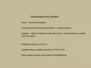
Organization • Each tract mediates specific modalities of sensation, somatotopic organization in tracts and cortex • Mechanoreceptive • Mechanical displacement of nerve endings • Touch (fine and diffuse), pressure, vibration, kinesthesia • Thermoreceptive • Cold and Heat • Nociceptive • Pain
Specialized Receptors • Receptors specialize by type of stimulus • Adaptiveness: Reduction of response to sustained stimuli • Basic Types of Sensory Receptors • Encapsulated Endings • Adapting (tactile) • Pacinian corpuscle: deep pressure touch and high frequency vibration. • Meissner’s corpuscle: light touch, such as the fingertips, palms, soles, lips, tongue, face • Free Nerve Endings (pain, temp, some tactile) • Nonadapting • Expanded Tip Endings (tactile, temp) • Moderately adapting
Three neuron Organization • 1st Order • Dorsal Root Ganglion • 2nd Order • Enter CNS at spinal cord or brainstem • Project to opposite side crossing midline to thalamus • 3rd Order • Thalamus neurons which project to cortex Dorsal root ganglion (‘spinal ganglion’)
Discriminative Touch Cerebral Cortex Bipolar or multipolar 3 Thalmus 2 Dorsal root ganglion Medulla Receptors (skin, muscle, joints) 1 Spinal cord Pseudo-Unipolar nerve
Anatomical Divisions • Dorsal Column-Medial Lemniscal (or Epicritic System) • Fine discriminative touch, vibration, limb position, kinesthesia & deep pressure • Position sense • Proprioception - Awareness of limb position • Kinesthesia - Awareness of limb movement • Anterolateral (or Protopathic System) • Pain, temperature and diffuse touch • Lateral spinothalamic tract • Anterior spinothalamic tract • Dorsal Column-Medial Lemniscal System
Dorsal Column-Medial Lemniscal System • Important for skilled movements • Stereognosis - Fine touch discrimination • Graphesthesia - Recognizing numbers written on body • Two and multiple point touch • Deep touch • Receptors • Meissner’s and Pacinian Corpuscles • Encapsulated end receptors • Highly sensitive and adaptable • Muscle Spindle Organs • Kinesthesia • Proprioception
Neural Pathways • Fasciculus Gracilis (slender, graceful) • Fasciculus Cuneatus (wedge-shaped – short) (think cuneiform writing) • Path • Spinal Ganglion (1) • Fasciculus Gracilis/Cuneatus tracts (1) • Gracilis or Cuneatus Nucleus (2) • Through Medial Lemniscus to Thalamus (3) • Thalamus to Cortex Mediate discriminative Touch from different Body areas; follow three-neuron organization
Levels of Reception • Fasciculus Gracilis • Sacral to Midthoracic Level • Lower Body • Fasciculus Cuneatus • Above Midthoracic Level • Upper Body
Pathway • Spinal Cord • Brainstem Nuclei • Thalamus (N. Ventral Posterolateralis) • Thalamus through Internal Capsule to Primary Sensory Parietal Cortex • Primary to Association Cortex • Mapped spatially (homunculus)
Dorsal Column-Medial Lemniscal System • In the PNS/Spine Pacinian corpuscle Cervical Thoracic Lumbar Sacral Fasciculus cuneatus Fasciculus gracilis Meissner’s corpuscle
Dorsal Column-Medial Lemniscal System Pons and Medulla Nucleus gracilis (lower body) Nucleus cuneatus (upper body) Medulla Decussation
Dorsal Column-Medial Lemniscal System • Midbrain-Cortex Homonculus Thalamus Midbrain Medial lemniscus
The homunculus (little man) • The motor strip (red, frontal cortex) spatially map corresponding portions of the contralateral hemisphere.
Clinical Considerations • If injury is inferior to decussation, deficit can be ipsilateral (same side) • If injury is superior to decussation, deficit will be contralateral (opposite side) • Tests • Two Point Discrimination • Stereognosis: ID object with eyes closed • Graphesthesia: number or letter on skin • Vibratory: Tuning fork on bony surface • Romberg: standing with eyes closed • Kinesthesia: movement identified • Association: Identification of object
Anterolateral system • Pain, Temperature, & Diffuse Touch • Three-tier neuron organization system • Enter at spinal ganglion (1st) • Cross in spinal tract (2nd order) • Ventral posterolateral n. of thalamus (3rd) • Two Tracts • Lateral Spinothalamic • Anterospinothalamic
Lateral Spinothalamic Tract • Receptors - Free Nerve Endings • Neural Pathway • Nocioceptors (pain) • Dorsolateral spinal cord (up or down several segments) • spinal cord entrance • Substantial Gelatinosa and Proprius • Where 1st order neurons connect with 2nd order neurons • Lateral Spinothalamic Tract • Cross Midline (2nd order) • Spinal Lemniscus (brainstem) • Thalamus (VPL) to Cortex • Collaterals to Subcortical structures
Pain and Temperature (antero-lateral) Bipolar or multipolar Cerebral Cortex 3 Dorsal root ganglion Thalmus 2 Receptors (skin, muscle, joints) 1 Spinal cord Pseudo-Unipolar nerve
Clinical Considerations (lesion locations) • PNS or spinal before midline cross results in problems ipsilaterally. • Spinal or Brainstem lesion results in problems contralaterally. • Chordotomy (surgical lesion) to reduce pain • Dermatomes: Failure to perceive pain
Dermatome • Dermatome: Refers to the body area innervated by the neurons in a single dorsal root ganglion (dorsal part of the spinal nerve)
Dermatome • Can help distinguish between psychiatric and neurological injury. • Psychiatric conversion disorder: often glove/stocking anesthesia • Neurological disorder: follows dermatomes
Other Considerations • Referred pain: one site has pain but felt in another site • Drugs can suppress pain sensitivity or block pathway • Analgesia: No sensation • Hypalgesia: Decreased pain (higher threshold) • Hyperalgesia: Increased pain (lower threshold)
Anterospinothalamic Tract • Discrimination of Diffuse touch • Receptors: All three types • Encapsulated endings • Free nerve endings • Expanded tip endings • Neural Pathway • Skin to ganglia (1st) • Dorsolateral spinal cord (up and down seg) • Proprius and Substantia Gelatinosa (2nd) • Go to spinothalamic tract to VPL (thalamus) to postcentral gyrus • Collaterals to subcortical structures • Clinically, interruption causes no obvious deficit
Collaterals in the axon Cortex VPL in thalamus Subcortical structures
Sensation from the head • Face and Head area • face • forehead • anterior half of scalp • dura mater • orbital cavities • nasal and oral cavities • Epicritic (Dorsal) and Protopathic (Anterolateral) Systems
Facial sensation • Three Neuron Levels • 1st order: Semilunar ganglion of Trigeminal Nerve • 2nd order: Principal sensory nucleus and trigeminal spinal tract nucleus • 3rd order: VPL in thalamus to lower third of postcentral gyrus
Fine Discriminative Touch • Neural Pathway • Encapsulated receptors in facial and head skin • Semilunar ganglion and trigeminal nucleus • Medial Lemniscus Thalamus to cortex
Cranial Proprioceptive and Kinesthetic Sensation • Teeth, periodontium palate, TMJ, muscles of mastication • Involves mesencephalic N. and follows similar pattern • Mechanism for jaw reflex and bit control
Cranial Sensation: Clinical Considerations • Lesions can affect only one branch • Ophthalmic • Maxillary • Mandibular • Or one half of the face • Tests the same for discrimination
Pain and Temperature from Face • Neural Pathway • Nocioceptors • Semilunar ganglion to • nucleus of spinal trigeminal tract (moves caudally) • chief sensory nucleus • Cross midline to thalamus and some stay ipsilateral • Postcentral Gyrus
Trigeminal Cranial Nerve Cerebral Cortex 3 Thalmus 2 1 Brainstem Spinal Cord
Clinical Considerations • Inflammation of semilunar ganglion causes severe pain • Tic douloureux - severe pain • Assessment of normal function • pinching to cause pain • Quality assessment by patient
Diffuse Touch from Face • Neural Pathway • Dorsal and ventral secondary trigeminal tract • Some to spinal trigeminal tract nucleus • Some to chief sensory nucleus • To ventral posteromedial nucleus of thalamus • To sensory cortex
Unconscious Proprioception • Conscious proprioception by dorsal column-medial lemniscal system • Unconscious involved in walking, articulating, writing, swallowing, and eye movement. • Two order neural system • Tracts • Dorsal Spinocerebellar • Cuneocerebellar • Ventral Spinocerebellar • Receptors • Muscle spindles and Golgi tendon organs located in muscles and limb joints
Ventral Spinocerebellar Tract • Mediates unconscious proprioception • Lower limbs to bilateral cerebellum • Sacral and Lumbar levels through ventrolateral Spinocerebellar tract to opposite cerebellar hemisphere
Dorsal Spinocerebellar Tract • Mediates unconscious proprioception • Lower limbs and middle regions of body to to bilateral cerebellum • Spinal ganglion to nucleus dorsalis of Clark at third lumbar segment • Do not cross and enter ipsilateral cerebellar hemisphere Dorsal spinocerebellar tract – information about movement (sensory feedback) Ventral spinocerebellar - internally generated information about the movement.
Cuneocerebellar Tract • Mediates upper limbs and neck • Uncrossed fibers to ipsilateral external cuneate nucleus to cerebellum • Clinical Considerations • Romberg used to determine some function • Difficult to test clinically Romberg Test Ask individual to stand straight with feet together and hands by the sides. Compare balance with eyes open versus eyes closed. If less steady with eyes closed (positive), ataxia is sensory – spinal injury. If there is no difference (negative) it suggests cerebellar problem.
MCQ • Which is the nucleus? • A • B • C • D A B C D
MCQ • Which is the node of ranvier? • A • B • C • D A B C D
MCQ • Which is the nucleus? • A • B • C • D A B C D