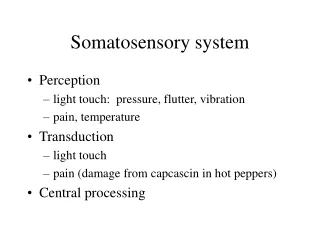
Somatosensory System Touch - mechanoreception
450 likes | 707 Vues
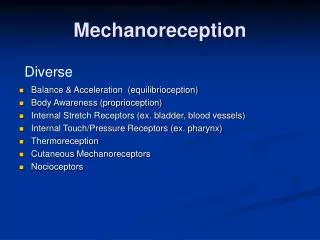
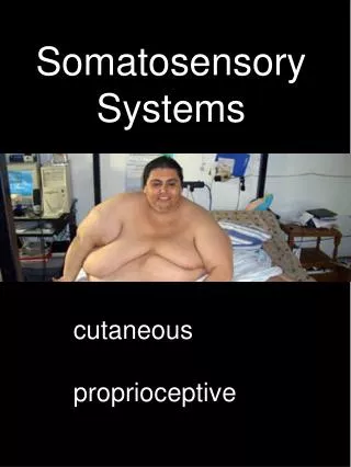
Somatosensory System Touch - mechanoreception Limb posture movements and forces – proprioception ( Haptics – object recognition through touch + proprioception , usually w ith the hand) Readings: Squire et al Ch 25 Supplementary reading: Kandel et al Chs 22- 23

Somatosensory System Touch - mechanoreception
E N D
Presentation Transcript
Somatosensory System Touch - mechanoreception Limb posture movements and forces – proprioception (Haptics – object recognition through touch + proprioception, usually with the hand) Readings: Squire et al Ch 25 Supplementary reading: Kandel et al Chs22-23 http://www.youtube.com/watch?v=FKxyJfE831Q
Somatosensory perception and action tightly linked. The somatosensory pathways of the brain integrate information from thousands of sensors in each hand and transforming it to a form suitable for cognition.ie force on a tool is located at the contact point of the tool by monitoring the vibrations and forces produced by those distant conditions. Also complex object recognition from spatio-temporal signatures. Sensory information is extracted for the purpose of motor control as well as cognition, and different kinds of information are extracted for those purposes. We can, for example, shift our attention from the baseball’s shape to its location in the hand to readjust our grip for an effective throw. This selective attention to different aspects of the sensory information is brought about by cortical mechanisms. Need to distinguish between active and passive touch: Motor deficits such as weakness, stiffness, or clumsiness may result from sensory loss, which is why passive sensory testing is important in the neurological examination.
+ Hair follicle afferents Most Sensitive to deformation 20-50 corpuscles/afferent ?? RA1 SA2 PC (RA2) SA1 Slipping object/large rf’s High density Form/braille Texture-hardness roughness Transmitted vibration textures Stretch?
Reaction to slippage: 65 msec - 45 msec is peripheral nerve and motor response time. Applied forces in response to shape of grasped object - 100 msec.
Dorsal root ganglion cells serve a dual role of transduction and information transmission. Multiple fibers from the axon branch to form a large receptive field. Receptive fields in the skin overlap.
Large variation in two-point threshold across the body surface The Spatial Resolution Varies Because the Density of Mechanoreceptors Varies
RA mechanoreceptor responds to sinusoidal mechanical stimuli with a single action potential for each cycle. The lowest stimulus intensity that evokes one action potential per cycle of the sinusoidal stimulus is called the receptor's “tuning threshold.” Merkel: 5-15 Hz Meissner: 20-50 Hz Pacinian: 60-400 Hz 25 Hz vibratory stimulus Tuning thresholds for vibration Compare with vision RA’s – lower thresholds
Variation in receptive field size for different receptor types
Spatial characteristics of stimuli depend on activity across population of receptors.
merkel meissner ruffini pacinian
Stimulus intensity in encoded by spike rate Neural response Perceived magnitude (pressure)
The muscle spindle is located within skeletal muscle and is excited by stretch of the muscle. It consists of a bundle of thin (intrafusal) muscle fibers entwined by a pair of sensory axons, and is also innervated by several motoraxons (not shown) that produce contraction of the intrafusal muscle fibers. Stretch-sensitive ion channels in the sensory nerve terminals are linked to the cytoskeleton by the protein spectrin
B. The depolarizing receptor potential recorded in a group la fiber innervating the muscle spindle (upper record) is proportional to both the velocity and amplitude of muscle stretch parallel to the myofilaments (lower record). When stretch is maintained at a fixed length, the receptor potential decays to a lower value. Although the receptor potential and firing rates of the sensory axons are proportional to muscle length, these responses can be modulated by higher centers in the brain that regulate contraction of intrafusal muscles. In this manner the spindle afferents are able to signal the amplitude and speed of internally generated voluntary movements as well as passive limb displacement by external forces.
Proprioceptors in muscle spindles and tendons Proprioceptors Ia, Ib, and II
Golgi tendon organs, located between skeletal muscle and tendons, measure the forces generated by muscle contraction. Although these receptors play an important role in reflex circuits modulating muscle force, they appear to contribute little to conscious sensations of muscle activity. Psychophysical experiments in which muscles are fatigued or partially paralyzed have shown that perceived muscle force is mainly related to centrally generated effort rather than to actual muscle force. Joint receptors play little if any role in postural sensations of joint angle. Instead, the perception of the angle of proximal joints such as the elbow or knee depends on afferent signals from muscle spindle receptors and efferent motor commands. Likewise, conscious sensations of finger position and hand shape depend on stretch receptors in the skin as well as muscle spindles and possibly joint receptors.
All four mechanoreceptors detect hand contact with the object but each monitors a different aspect of the action as the task progresses. SA1 fibers encode the grip force and SA2 fibers the hand posture. RA1 fibers encode the rate of force application and movement of the hand on the object. RA2 fibers sense vibrations in the object with each movement: at hand contact, lift-off, table contact, and release of grasp.
dorsal column-medial lemniscalsystem for tactile sensations and proprioception and anterolateral system for pain and temperature
The fibers of the medial lemniscus, (tactile and proprioceptive signals), terminate in the ventral posterior nucleus of the thalamus. The medial zone of the nucleus receives trigeminal nerve fibers from the head and face. The lateral zone receives fibers from the dorsal column nuclei; these inputs are arranged somatotopically, with the forelimb medial and the trunk and legs lateral. The ventral posterior nucleus— called nucleus ventraliscaudalis in humans— has traditionally been thought of as a single nucleus in which the fibers carrying cutaneous signals terminate in a large central and caudal region while those conveying proprioceptive information terminate in a dorsal and rostral region. More recently, Jon Kaas and his colleagues have argued that the two regions represent separate nuclei: the ventral posterior nucleus proper, which receives the cutaneous information conveyed by medial lemniscal and trigeminal axons, and the ventral posterior superior nucleus, which processes proprioceptive information. These nuclei send their outputs to different subregions of the cerebral cortex. The ventral posterior nucleus transmits cutaneous information primarily to area 3b of the primary somatosensory cortex, whereas the ventral posterior superior nucleus conveys proprioceptive information principally to area 3a in the postcentralgyrus.
Serial + parallel processing Proprioceptive input
Dorsal path to superior parietal lobule, providing areas in that lobule with somesthetic information for control of voluntary movements, selective attention, and information about how to perform different tasks, sometimes known as the “how” pathway. Ventral path to SII and from there to the caudal insula, to areas of the temporal lobe, and to premotor and prefrontal cortical areas. Eventual convergence of somatosensory, auditory, and visual information in the medial temporal lobe is viewed as the path toward shape and form processing. This ventral path is vital for inserting new information into declarative memory and accessing established memories for comparison with ongoing events.
Tactile information from the fingertips is used faster than can be readily explained by rate codes. (Johansson & Birznieks NN 2004) Reaction to slippage: 65 msec - 45 msec is peripheral nerve and motor response time. Applied forces in response to shape of grasped object - 100 msec.
Excitatory and inhibitory zones of receptive fields of neurons in area 3b. The excitability of cortical neurons varies over time and as a function of the stimulus location on the skin. Peak excitation occurs in the middle of the receptive field 15 to 25 ms following brief taps on the skin. Inhibition occurs shortly thereafter and is strongest at 45 ms. Delayed inhibition allows each stimulus to be perceived as a distinct event when delivered at rates lower than 25 Hz (cf vision). Each map indicates the intensity of excitation (red) and inhibition (blue) produced over 10 ms periods following brief taps to a small patch of skin on the fingertip. Cortical neurons vary in the relative strength and spatial location of excitatory and inhibitory fields.
Velocity invariance of cell’s responses.
Receptive fields of neurons in S1 are larger than those of the sensory afferents. Each of the hand figurines shows the receptive field of an individual neuron in areas 3b, 1, 2, and 5, based on recordings in alert monkeys.Colored regions indicate the region where light touch elicits action potentials. Neurons at later stages of cortical processing (areas 1 and 2) have larger receptive fields and more specialized inputs than neurons in area 3b. The neuron from area 2 is directionally sensitive to motion toward the fingertips. Neurons in area 5 often have symmetric bilateral receptive fields at mirror image locations on the contralateral and ipsilateral hand. Ablation of area 3b: general losses Ablation of area 1: loss of texture discrim Ablation of area 2: loss of 3D form discrim Direction sensitivity emerges in Area 1 and 2, not present in 3b
Note overrepresentation of Hand, etc and Underrepresentation of Torso Cortical magnification Similar to vision.
Task dependent responses in S-ii but not S-I In an elegant study RanulfoRomo and his colleagues compared responses of neurons in S-I, S-II, and various regions of the frontal lobe of monkeys while the animals performed a two-alternative forced-choice task . The animals were rewarded if they correctly recognized which of two vibratory stimuli was higher in frequency. Neurons in S-I faithfully represented the vibratory cycles each stimulus, firing a brief burst in response to each. In contrast, S-II neurons responded to the first stimulus with spike trains proportional to its frequency but responded to the second stimulus with a signal that combined the frequencies of both stimuli. Thus the same vibratory stimulus could evoke different firing rates in S-II, depending on whether the preceding stimulus was higher or lower in frequency.
During reaching and grasping, neural activity in the posterior parietal cortex coincides with activation of neurons in motor and premotor areas of the frontal cortex and precedes activity in S-I. There is strong evidence that area 5 receives convergent central and peripheral signals that allow it to compare central motor commands with peripheral sensory feedback during reaching and grasping behaviors. Sensory feedback from the hand to the posterior parietal cortex is used to confirm the goal of the planned action, thereby reinforcing a previously learned skill or correcting those plans when errors occur. Predicting the sensory consequences of hand actions is an important component of active touch. For example, when we view an object and reach for it, we predict how heavy it should be and how it should feel in the hand; we use such predictions to initiate grasping. Corollary discharge from motor areas to somatosensory regions of the cortex may play a key role in active touch. It provides posterior parietal neurons with information on intended actions, allowing these neurons to compare planned and actual neural responses to tactile stimuli.
Orientation tuning in SII receptive fields. Finger pads have similar tuning. Results from lateral inhib, not composition of inputs as in vision Ablation of SII: Loss of discrim of shape and texture Doesn’t affect SI responses Ablation of SI silences SII Responses modulated by attention
Two patients with lesions to the anterior parietal lobe show severe impairment in both sets of tactile tests but only moderate impairment in the motor tasks. Touch, discrimination Shape recognition Grip force Skilled movement Eg peg in slot. Three patients with posterior parietal lesions show only minor deficits in simple somatosensory tests but severe impairment in complex tests of stereognosis and form. Motor deficits are greater in skilled tasks. . Four patients with combined lesions to anterior and posterior parietal cortex show severe impairment in all tests. Interestingly, the patient who showed the least impairment in this group (patient f) suffered brain damage at birth; the developing brain was able to compensate for the loss of major somatosensory areas. Lesions in the other patients resulted from strokes later in life.
Effect of lesion in area 2 - loss of ability to coordinate fingers, plus sensory deficits.
Mechanoreceptors Differ in Morphology and Skin Location RA SA1 SAII